

Using cholangiocyte organoids to regenerate human bile ducts
Posted: 22 June 2021 | Dr Fotios Sampaziotis (University of Cambridge) | No comments yet
A team of researchers has shown that injection of cholangiocyte organoids in human livers ex vivo can repair the organs’ bile ducts. In this article, Dr Fotios Sampaziotis explains how his team’s study provides the first proof-of-principle for the efficacy of cellular therapies using organoids in human.
The natural history of most diseases is cumulative organ damage, leading to progressive deterioration and ultimately organ failure. At this terminal stage, the only effective therapy is transplantation. This is particularly pronounced for liver disorders, which constitute the leading cause of death in 35-49 year olds and are one of the top causes for mortality regardless of aetiology.1,2 Effective treatments are available only for certain indications,3,4 rendering liver the second-most commonly transplanted organ.5 However, this approach is restricted by organ availability, complications related to the operation or the need for long‑term immunosuppression and cost.6 Seven percent of patients on the waiting list die before they receive a graft,7 while mortality from liver disease continues to increase over time.2 Therefore, new therapeutic options are urgently needed.
Cellular therapies hold great promise for addressing these challenges.8 Administering healthy cells capable of repairing and regenerating damaged organs to patients provides a unique alternative to liver transplantation. These cells can be grown in almost unlimited supply, overcoming issues related to graft availability. They are technically easier to administer and they are associated with less complications, while being at a fraction of the cost compared to liver transplantation. Furthermore, cellular therapies can be administered to a broader number of patients compared to liver transplantation. These include individuals with early disease, which is refractory to alternative treatment or patients with very advanced disease who are not adequately fit to survive transplantation. Finally, autologous cells may be used, overcoming the need for long‑term immunosuppression, while disease recurrence can be addressed with repeated administration of cells.
Despite these advantages, the translation of cell-based therapy into clinical products has been hindered by the lack of data demonstrating the efficacy and safety of this approach in human. Indeed, the number of ongoing clinical trials is restricted, while animal studies are limited by interspecies variation.
A focus on the bile duct
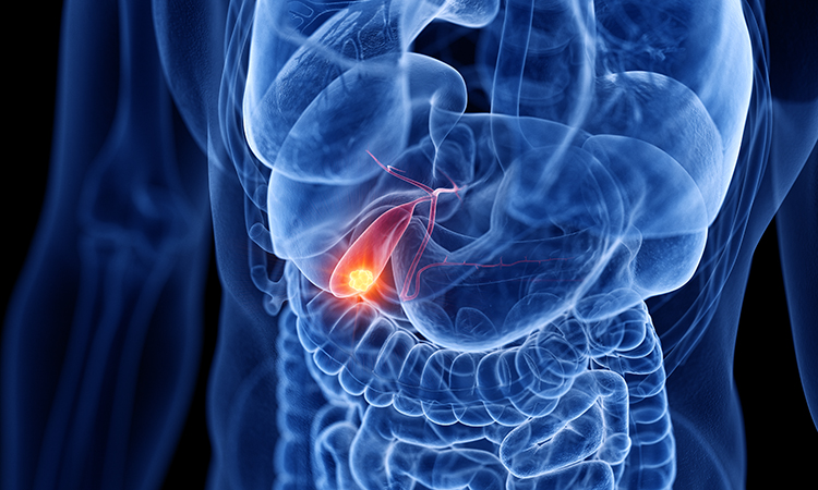
We decided to focus on the bile duct as a proof-of-principle system to address these challenges for two reasons. Firstly is their clinical impact; the biliary tree forms a tubular network in the liver transferring bile to the duodenum. Bile duct disorders (cholangiopathies) carry significant morbidity and mortality. They affect children or young adults and account for 25 percent of adult and 70 percent of paediatric liver transplantations,4,9 with no therapeutic alternatives for advanced disease.9,10 Importantly, liver transplantation is not always curative. Disease recurrence or other biliary complications occur in up to a third of these patients.4 Secondly, biliary epithelial cells (cholangiocytes) constitute an optimal cell type for cellular therapies. They exhibit a unique regeneration potential11,12 and may even act as facultative stem cells in certain animal models.13 Furthermore, biliary tissue is one of the most abundantly available and easily accessible tissues because gallbladder excision (cholecystectomy) is the most common operation worldwide,14 while cholangiocytes can also be obtained through minimally-invasive procedures such as Endoscopic Retrograde Cholangiopancreatoraphy (ERCP) or liver biopsies.15
The development of bile duct organoids
To grow cholangiocytes for cellular therapy, we decided to start from primary biliary tissue. Primary cells provide an optimal starting material for regenerative medicine applications because they already exhibit the required markers and function to perform their physiological role. Furthermore, they are associated with an optimal safety profile, as they do not require genetic manipulation, while using autologous cells can further minimise risks and expedite regulatory approval. Nevertheless, when we started our research, the use of primary cells was hampered by loss of function and reduced proliferation in culture. We discovered that to maintain their function, cholangiocytes needed to be organised in structures with a central lumen resembling bile ducts. To achieve this, we grew the cells in three-dimensional hydrogels, which allowed them to self-organise into hollow-lumen organoids and incorporate key components of the cells’ native niche in our culture. Under these conditions, cholangiocytes maintained their function and proliferative capacity.16
To explore the potential of these cholangiocyte organoids for regenerative medicine, we first decided to assess how similar these organoids were to the cells they would be replacing. To achieve this, we compared cholangiocyte organoids in culture to their native counterparts. We observed small differences in cholangiocytes in vivo versus in vitro, however we also discovered that cholangiocytes are plastic. These differences were a result of the cells adapting to culture and they were reversed when our organoids were exposed to their native niche.17 To validate these findings and confirm the regenerative potential of our organoids, we transplanted cholangiocyte organoids into immunocompromised mice with diffuse cholangiopathy. Our results showed that the cells engrafted in the intrahepatic branches of the biliary tree, repaired damaged ducts and rescued the animals.17 To explore whether these findings can be translated in human, we then used human liver grafts, which were perfused ex vivo and were deemed not suitable for transplantation. We injected our cells in the biliary tree of these organs and demonstrated for the first time that cholangiocyte organoids can repair bile ducts and restore their function in human.17
The most promising findings
One of the most promising findings of the study was the discovery that cholangiocyte organoids are plastic and that their identity is controlled by their niche.17 This implies that any cholangiocyte can give rise to the same organoids and therefore, cholangiocytes are interchangeable for regenerative medicine applications. Furthermore, our study provides the first demonstration for the efficacy of cellular therapies in the context of bile duct disorders and the first proof-of-principle that human organoids from any tissue can repair human organs. Finally, our results pioneer a novel regenerative medicine approach – the use of cell‑based therapy to repair marginal donor organs ex vivo, render them suitable for transplantation, increase organ availability and reduce pressure on the liver transplant waiting list.
Challenges faced
To achieve these results, we had to overcome several practical limitations. Cholangiocytes represent only three percent of liver parenchyma cells; therefore their transcriptional signature is lost in bulk analyses of liver tissue. Using cell sorting is possible, but this method still pools together all cholangiocytes and does not enable different populations to be discerned. To overcome these limitations, we used single-cell RNA sequencing. The data we obtained allowed us to not only characterise cholangiocyte populations in organoid and primary tissue at the highest possible resolution, but also to use this information to identify mechanisms controlling cholangiocyte identity.
Furthermore, cellular therapies may be limited by poor engraftment efficiency, escape of cells in the system circulation and heterotopic cell engraftment. To overcome these issues and ensure that transplanted cells engrafted in the intended site, we took advantage of the fact that the bile ducts constitute a blind-ended tubular network, with only one outflow, the Common Bile Duct (CBD). We developed a method for injecting cholangiocyte organoids directly in bile ducts and blocking the outflow of bile for a limited amount of time to promote engraftment.
Finally, one of the biggest challenges was assessing the efficacy of cholangiocyte organoids as a cellular therapy in human. To achieve this, we used human livers receiving ex vivo normothermic perfusion (EVNP). This approach is routinely used in the context of liver transplantation in our hospital to maintain liver grafts in near‑physiological conditions, by circulating warm oxygenated blood through the organ and to assess graft function prior to transplantation.18,19 We captured livers which were deemed unsuitable for transplantation due to duct damage, evidenced by changes in the composition of bile. To maintain the number of cells required for the injection to a minimum, we used radiological guidance to inject our cells in a peripheral branch of a bile duct, with only a small area of distribution. This allowed us to generate the number of organoids required to regenerate the selected area.
The future of organoids in cell therapies
In conclusion, our study illustrates the key role of the niche in controlling cholangiocyte organoid identity and raises the possibility that similar mechanisms may apply in other epithelial organoids. In parallel, it provides the first proof‑of‑principle for the efficacy of regenerative medicine using organoids in human. We expect this approach to be used by multiple other groups to validate different types of organoids and expedite regulatory approval of these cells for first‑in‑human trials. Finally, our findings pioneer a novel regenerative medicine strategy, repairing donor organs ex vivo so that they can be used for transplantation.
About the author
Dr Fotios Sampaziotis is a physician hepatologist in the Cambridge University Hospitals Liver Unit and a Clinical Lecturer in the University of Cambridge. He is the co-founder and CEO of Bilitech LTD, which aims to advance innovative cellular and bioengineered therapies to clinical products, and a founding member of the European Association for the Study of Liver consortium for Regenerative Hepatology. His work on regenerative medicine, bioengineering and hepatobiliary disease modelling has received multiple awards, including the Medawar Medal, Dame Sheila Sherlock award and the UK National Scholar award. Fotios earned his medical degree from the University of Athens and his PhD in regenerative medicine from the University of Cambridge.
References
- Office for National Statistics – NOMIS OLMS. Mortality Statistics – underlying cause sex and age. http://www.nomisweb.co.uk/articles/1128.aspx. 2019.
- British Liver Trust. The alarming impact of liver disease in the UK. 2019;298858:1–37.
- Gallo A, Esquivel CO. Current options for management of biliary atresia. Pediatr Transplant [Internet]. 2013;17(2):95–8. Available from: http://doi.wiley.com/10.1111/petr.12040
- Enestvedt CK, Malik S, Reese PP, et al. Biliary complications adversely affect patient and graft survival after liver retransplantation. Liver Transpl [Internet]. 2013 Sep [cited 2020 Apr 25];19(9):965–72. Available from: http://www.ncbi.nlm.nih.gov/pubmed/23818332
- NHS Blood and Transplant. Overview of Organ Donation and Transplantation. Organ Donation Transplant Act Rep 2014/15. 2015;(March 2018):4–11.
- Asrani SK, Harshad Devarbhavi, Eaton J, Kamath PS. Burden of liver diseases in the world. J Hepatol. 2019;70(1):151–71.
- NHS Blood & Transplant. Liver Transplant Activity Report. 2020;(February):77–88.
- Kimbrel EA, Lanza R. Next-generation stem cells — ushering in a new era of cell-based therapies. Nat Rev Drug Discov. 2020;19(7):463–79.
- Murray KF, Carithers RL. AASLD practice guidelines: Evaluation of the patient for liver transplantation. Hepatology [Internet]. 2005;41(6):1407–32. Available from: http://doi.wiley.com/10.1002/hep.20704
- Leithead JA, Ferguson JW. Chronic kidney disease after liver transplantation. J Hepatol [Internet]. 2015 Jan [cited 2017 Aug 19];62(1):243–4. Available from: http://www.ncbi.nlm.nih.gov/pubmed/25263005
- Lu WY, Bird TG, Boulter L, et al. Hepatic progenitor cells of biliary origin with liver repopulation capacity. Nat Cell Biol. 2015;17(8):973–83.
- Rodrigo-Torres D, Affò S, Coll M, et al. The Biliary Epithelium Gives Rise to Liver Progenitor Cells Daniel. Hepatology. 2014;60(4):1367–77.
- Raven A, Lu WY, Man TY, et al. Cholangiocytes act as facultative liver stem cells during impaired hepatocyte regeneration. Nature. 2017;547(7663):350–4.
- NICE. Costing statement: Gallstone Disease. Implementing NICE guideline on gallstone disease (CG188) [Internet]. NICE Clinical Guideline 186. 2014 [cited 2018 Jan 4]. Available from: https://www.nice.org.uk/guidance/cg188/resources/costingstatement-pdf-193298365
- Tysoe OC, Justin AW, Brevini T, et al. Isolation and propagation of primary human cholangiocyte organoids for the generation of bioengineered biliary tissue. Nat Protoc. 2019 Jun 1;14(6):1884–925.
- Sampaziotis F, Justin AWAW, Tysoe OCOC, et al. Reconstruction of the mouse extrahepatic biliary tree using primary human extrahepatic cholangiocyte organoids. Nat Med [Internet]. 2017 Jul 3 [cited 2017 Aug 20];23(8):954–63. Available from: http://www.ncbi.nlm.nih.gov/pubmed/28671689
- Sampaziotis F, Muraro D, Tysoe OC, et al. Cholangiocyte organoids can repair bile ducts
after transplantation in the human liver. Science. 2021;371(6531):839–46. - Watson CJE, Kosmoliaptsis V, Pley C, et al. Observations on the ex situ perfusion of livers for transplantation. Am J Transplant [Internet]. 2018 Aug 1 [cited 2020 Apr 8];18(8):2005–20. Available from: http://www.ncbi.nlm.nih.gov/pubmed/29419931
- Nasralla D, Coussios CC, Mergental H, et al. A randomized trial of normothermic preservation in liver transplantation. Nature [Internet]. 2018 May 18 [cited 2018 Sep 5];557(7703):50–6. Available from: http://www.nature.com/articles/s41586-018-0047-9
Related topics
Biologics, Biopharmaceuticals, Cell Regeneration, Organoids, Regenerative Medicine
Related conditions
Cholangiopathies
Related organisations
OXGENE, Takara Bio