


news
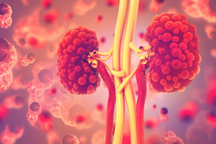
Tropical frog models reveal novel kidney disease insights
Scientists utilised CRISPR technology and deep learning systems to investigate the genes associated with polycystic kidney disease.
List view / Grid view
Scientists utilised CRISPR technology and deep learning systems to investigate the genes associated with polycystic kidney disease.

A new high-resolution virtual microscopy technique enables the rapid visualisation of tissue, paving the way for histopathology analysis during surgery.
A new computer-aided tool maps allosteric sites in G protein-coupled receptors to search for allosteric drugs to treat a range of diseases.
This whitepaper overviews phenotypic and functional characterisation of CAR-T cells with advanced flow cytometry and live-cell analysis.
The statistical method known as maximum entropy could improve cryogenic electron microscopy (cryo-EM) for more effective drug treatments.
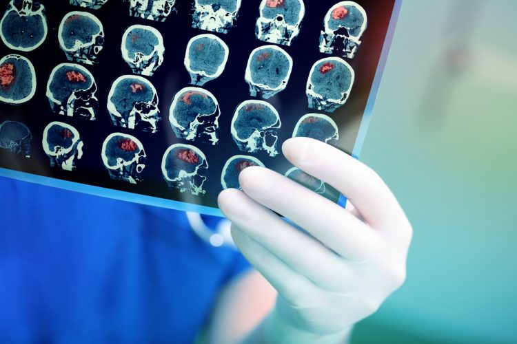

The Wyss Centre has announced a collaborative project to improve understanding of the brain cancer glioblastoma and develop new personalised therapies.

Researchers have used CRISPR and cryogenic electron microscopy to unravel the workings of two receptors involved in diseases such as cancer and COVID-19.
Researchers have developed an inexpensive method for visualising blood flow in the brain that can discern the motions of individual blood cells.
Evaluation of neurotoxicity effects is an active area of investigation in drug discovery and disease modeling.
This whitepaper describes several live-cell phenotypic analyses suitable for the characterisation of astroglia cells.
The cell painting assay uses up to six fluorescent dyes to label and visualize a variety of subcellular structures at the single cell level.

In this original report, find an in-depth analysis of AI and informatics within imaging, synthetic biology, drug screening and drug design. Featured interviews with experts from AstraZeneca, Auransa, PolarisQB and Chalmers University of Technology.
The latest edition of the live-cell analysis handbook is a companion guide for live-cell analysis users. Includes discussion of live-cell analysis.
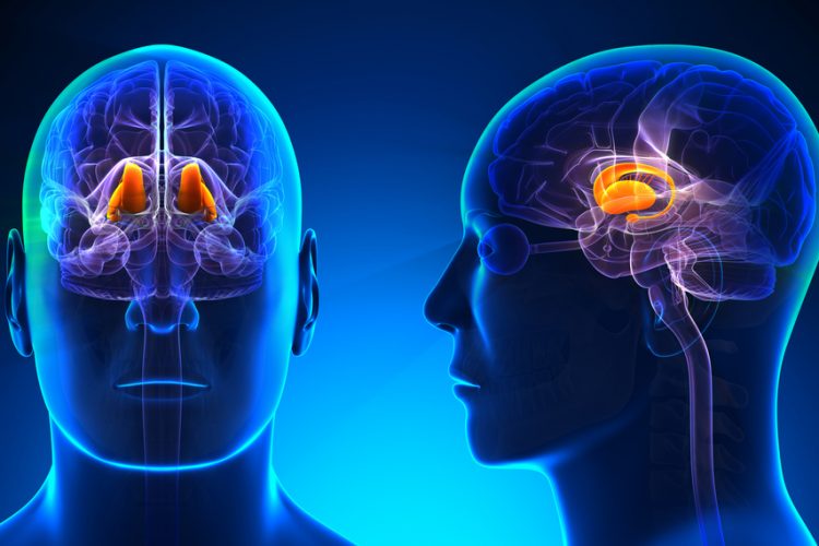
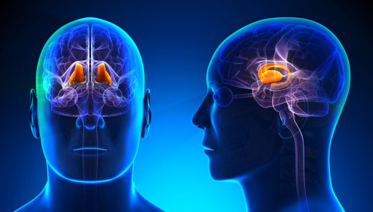
Research shows that cells gather more data inside the thalamus than once believed, potentially changing medicines for brain disorders.
Depletion of ATP due to viral-induced CPE leads to a reduction in luminescence signal, enabling quantitation of viral-induced CPE in host cells.