

Wyss Centre initiates collaboration to improve brain tumour treatment
Posted: 13 October 2021 | Anna Begley (Drug Target Review) | No comments yet
The Wyss Centre has announced a collaborative project to improve understanding of the brain cancer glioblastoma and develop new personalised therapies.
The Wyss Center, Switzerland, announced today that it will lead a collaborative project to improve understanding of the brain cancer glioblastoma (GBM) and help develop more effective therapies. The project, in collaboration with the University of Geneva, Switzerland, and the Paris Brain Institute, France, will map the GBM microenvironment in whole human tumours.
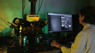
The collaboration will use light-sheet microscopy to image entire glioblastoma tumours in 3D without slicing or damaging the samples. [credit: Wyss Center for Bio and Neuroengineering]
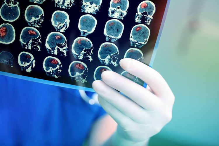
GBM is an incurable disease and is the most common and aggressive brain tumour worldwide. The tumour microenvironment (TME) includes tumour cells, immune cells, molecules released by these cells, blood vessels and hypoxic areas of low oxygen all of which change over time and may contribute to the recurrence of GBM. By accurately mapping the TME in recurrent tumours from the same patient, before and after treatment, the team will build a model that helps predict response to therapies.
According to the researchers, investigations are typically performed on very thin GBM slices, only about 10 microns thick, rather than on complete human tumours and so fail to capture the complexity of the TME in its entirety. In addition, standard analyses do not reveal the way that the microenvironment changes over time with treatment, nor the reasons why treatment might be failing.
Drug Target Review has just announced the launch of its NEW and EXCLUSIVE report examining the evolution of AI and informatics in drug discovery and development.
In this 63 page in-depth report, experts and researchers explore the key benefits of AI and informatics processes, reveal where the challenges lie for the implementation of AI and how they see the use of these technologies streamlining workflows in the future.
Also featured are exclusive interviews with leading scientists from AstraZeneca, Auransa, PolarisQB and Chalmers University of Technology.
“To address this challenge, we are bringing together a dynamic group of scientists. Our colleagues at the Paris Brain Institute are experts in tissue preparation and computational analysis and at the Wyss Center, we have deep expertise with fast and high-resolution light-sheet microscopy. We will look to understand the role of specific genes and proteins in the development of the tumour,” explained Stéphane Pagès, principal investigator at the Wyss Center.
By combining state-of-the-art molecular and cellular techniques, with advanced light-sheet imaging, the team will develop an innovative approach to map the TME. They will create three-dimensional (3D) maps of RNA sequences associated with specific genes thought to be involved in tumour development. They will also label specific proteins and use light-sheet microscopy to visualise and understand the interplay between cell types, blood vessels and hypoxic regions in the TME. Light-sheet microscopy will allow whole GBM tumours to be imaged with a sheet of light without damaging the sample.
ARTICLE: Telomerase – a universal cancer target for immunotherapeutic vaccines
The 3D maps will be integrated with clinical data from patients to understand how the disease progresses following therapy. The approach will be applied to tumours resected from two separate patient cohorts with recurrent GMB. One cohort consists of patients who are being treated with standard care – radio and chemotherapy. The other includes patients who have participated in an immunotherapy vaccine clinical trial.
“We hope that the unique insights of this ‘tissue time machine’ will mean that we can intervene in brain cancer earlier with a truly personalised approach based on the genetic profiles of individual tumours,” said Wyss Center’s Chief Executive Officer Mary Tolikas.
Related topics
chemotherapy, DNA, Drug Targets, Genetic analysis, Genomics, Imaging, Microscopy, Oncology, Personalised Medicine, Radiotherapy, RNAs, Therapeutics
Related conditions
brain cancer glioblastoma
Related organisations
Paris Brain Institute, University of Geneva, Wyss Center
Related people
Mary Tolikas, Stéphane Pagès