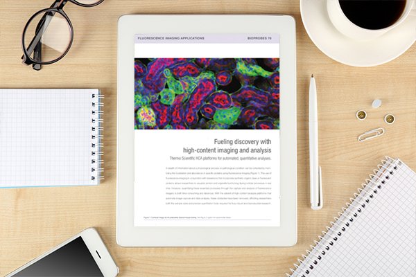
Application note: Fuelling discovery with high-content imaging and analysis
Posted: 10 May 2019 | Thermo Fisher Scientific | No comments yet
HCA platforms automate image capture and data analysis, giving researchers the sample sizes and precise quantitation tools required for research.
A wealth of information about a physiological process or pathological condition can be collected by monitoring the localisation and abundance of specific proteins using fluorescence imaging. The use of fluorescence imaging in conjunction with biosensors that incorporate synthetic organic dyes or fluorescent proteins allows researchers to visualise protein and organelle functioning during cellular processes in real time.
[pdf-embedder url=”https://www.drug.russellpublishing.co.uk/wp-content/uploads/5.-high-content-imaging-analysis-bioprobes-76-article-tracked-links.pdf” title=”Thermofisher – Bioprobes 76 PDF”]
Related content from this organisation
- Application note: Evaluation of hepatic function in 3D culture
- Application note: Hypoxia measurements in live and fixed cells
- Application note: High-throughput imaging and analysis of spheroids
- Application note: Oxidative Stress Measurements Made Easy
- Application note: Understanding Cell Death by High Content Analysis
Related topics
Drug Discovery, Screening, Stem Cells, Target molecule
Related organisations
High Content Analysis from ThermoFisher Scientific


