

Dynamics of GPCRs revealed using combination of approaches
Posted: 26 May 2020 | Hannah Balfour (Drug Target Review) | No comments yet
A team used both structural and spectroscopic techniques to study the dynamics of cell surface G-protein coupled receptors (GPCRs).
Authors of a new study demonstrated how combining structural and spectroscopic approaches enabled them to obtain precise information about functioning G-protein coupled receptors (GPCRs), the largest family of cell membrane receptors.
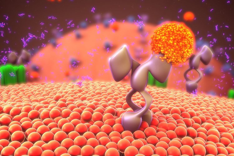
GPCRs are a family of receptors with seven transmembrane domains and couple to an intracellular protein, called a G protein, to relay a signal. Interaction of the signalling molecule with the receptor triggers a change in the three-dimensional (3D) structure of the receptor, which activates the G protein. The activated G protein triggers a signalling cascade inside the cell, which results in a response to the signal.
GPCRs are vital for a cell to survive and their disfunction has been implicated in neurodegeneration, cardiovascular diseases and cancer, and been shown to contribute to obesity, diabetes and mental disorders. This makes them a popular target for drugs; however, membrane receptor analysis has always been a slow and extremely laborious process that could not completely reveal the molecule’s behaviour inside the cell.
Drug Target Review has just announced the launch of its NEW and EXCLUSIVE report examining the evolution of AI and informatics in drug discovery and development.
In this 63 page in-depth report, experts and researchers explore the key benefits of AI and informatics processes, reveal where the challenges lie for the implementation of AI and how they see the use of these technologies streamlining workflows in the future.
Also featured are exclusive interviews with leading scientists from AstraZeneca, Auransa, PolarisQB and Chalmers University of Technology.
“Currently, scientists have two options when it comes to studying proteins. They can either ‘freeze’ a protein and have its precise static snapshot or study its dynamics at the cost of losing details. The former approach uses methods such as crystallography and cryogenic electron microscopy; the latter uses spectroscopic techniques,” said Anastasia Gusach, a research fellow at the Moscow Institute of Physics And Technology (MIPT) Laboratory of Structural Biology of G-protein Coupled Receptors, Russia.
The authors of a study published in Current Opinion in Structural Biology, used a combination of structural and spectroscopic techniques to study GPCRs and their function.
They found that the double electron-electron resonance (DEER) and the Förster resonance energy transfer (FRET) techniques act as an “atomic ruler”, ensuring precise measurements of distances between separate atoms and their groups within the protein. Nuclear magnetic resonance enabled them to visualise the overall shape of the receptor molecule, while modified mass spectrometry methods (MRF-MS, HDX-MS) indicated which parts of the molecule face outwards, based on its susceptibility to solvents.
“Studying the GPCR dynamics uses cutting-edge methods of experimental biophysical analysis such as nuclear magnetic resonance (NMR) spectroscopy, electron paramagnetic resonance (EPR) spectroscopy, and advanced fluorescence microscopy techniques including single-molecule microscopy,” said Alexey Mishin, deputy head of the MIPT Laboratory for Structural Biology of G-protein Coupled Receptors.
Gusach added: “Biophysicists that use different methods to study GPCRs have been widely organising collaborations that already bore some fruitful results. We hope that this review will help scientists specializing in different methods to find some new common ground and work together to obtain a better understanding of receptors’ functioning.”
A precise understanding of how GPCRs function and transfer between states would greatly expand the capabilities for structure-based drug design, according to the authors.
Related topics
Analysis, Analytical techniques, Disease research, Drug Targets, GPCRs, Molecular Targets, Protein, Proteomics, Structural biology, Technology
Related conditions
Cancer, cardiovascular diseases, Diabetes, Neurodegenerative diseases, Obesity
Related organisations
Moscow Institute of Physics and Technology (MIPT)
Related people
Alexey Mishin, Anastasia Gusach