

Novel label-free method enables imaging of metabolites
Posted: 10 February 2020 | Victoria Rees (Drug Target Review) | No comments yet
A label-free imaging technology has been developed by researchers, allowing them to investigate biomolecules such as metabolites, aiding in the study of drugs.
A novel label-free imaging method has been developed by a collaborative team from Helmholtz Zentrum München, Technical University of Munich (TUM) and Heidelberg University Hospital in Germany. According to the researchers, their technique will have a “major impact” on the basic understanding, monitoring, prevention and treatment of metabolic diseases, such as diabetes and obesity.
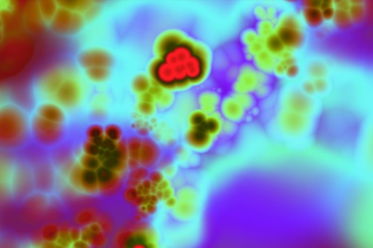
The new method provides real-time images of biomolecules in living cells without the need for labels or contrast agents. Called Mid-infraRed Optoacoustic Microscopy (MiROM), the technology allows researchers to induce specific molecular vibrations using mid-infrared lasers. Selective absorption of transient mid-infrared light from different molecules triggers a thermoelastic expansion, ie, a minute volumetric expansion that generates ultrasound waves. These are detected and processed to form an image of the distribution of specific molecules, depending on the wavelength(s) of excitation.
MiROM micrograph of living adipocytes (1mm x 1mm): lipids (red), proteins (green) and carbohydrates (blue) (credit: Helmholtz Zentrum München/Miguel A. Pleitez).
The researchers say that compared to previous techniques, one advantage of this new method is that it is not limited to dry-fixed samples. By detecting acoustic waves, which pass easily through tissue, rather than photons which are strongly absorbed by tissues and water, the process provides imaging features for metabolites beyond current technologies.
Drug Target Review has just announced the launch of its NEW and EXCLUSIVE report examining the evolution of AI and informatics in drug discovery and development.
In this 63 page in-depth report, experts and researchers explore the key benefits of AI and informatics processes, reveal where the challenges lie for the implementation of AI and how they see the use of these technologies streamlining workflows in the future.
Also featured are exclusive interviews with leading scientists from AstraZeneca, Auransa, PolarisQB and Chalmers University of Technology.
“MiROM offers a microscopy breakthrough. In conventional mid-infrared imaging, higher biomolecule concentration leads to higher signal loss. Conversely, MiROM converts mid-infrared imaging to a positive contrast modality, whereby higher concentration yields stronger signals. The technique demonstrated label-free imaging of biomolecules beyond the sensitivity limitations of Raman methods,” explained Professor Vasilis Ntziachristos, Director of the Institute of Biological and Medical Imaging and of the Chair of Biological Imaging at Helmholtz Zentrum München.
“MiROM provides novel insights into subpopulations of cells over time. Moreover, it enables us to detect not only lipids, but also carbohydrates and proteins in real-time,” said Miguel Pleitez, who led the system development.
In particular, the team say that MiROM can be used for the study of metabolites, to improve understanding of the molecular processes associated with fat storage and release due to lipolysis, the breakdown of white and brown fat cells. These insights could potentially be used in the development of treatments for diabetes or obesity. As previous methods could affect biological functions, readings from the new technology will improve imaging.
“MiROM offers unique in vivo label-free observations of metabolic processes in real-time, which can be applied to dynamically study the effects of different diets on the cellular level or evaluate the performance of new classes of drugs,” said Pleitez.
The team is now working on a revised version of MiROM with enhanced speed, resolution and sensitivity to boost discoveries in a broader spectrum of diseases including cancer.
Related topics
Imaging, Label-free, Metabolomics, Research & Development, Therapeutics
Related organisations
Heidelberg University Hospital, Helmholtz Zentrum München, Technical University of Munich (TUM)
Related people
Miguel Pleitez, Professor Vasilis Ntziachristos