

Protein imaging equipment enables enzymes to be observed in real-time
Posted: 19 November 2019 | Rachael Harper (Drug Target Review) | 1 comment
The imaging equipment, European XFEL, is said to mark a new age of protein movie-making and enables enzymes involved in disease to be observed in real-time.
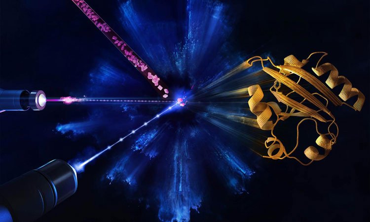
Microcrystals are injected (top, left) and a reaction is initiated by blue laser pulses hitting the proteins within the crystals (middle, left). The atomic structure of the protein (right) is probed during the reaction by the X-ray pulses (bottom, left). At the European XFEL, femtosecond optical laser pulses match the X-ray pulses that fire at a megahertz rate. X-ray pulses are six orders of magnitude larger than that at other X-ray sources. This makes it possible to produce diffraction patterns for nearly any protein, yielding still images recorded over rapid time increments that form molecular movies (credit: European XFEL/Blue Clay Studios).
Building on testing of the imaging equipment European X-ray Free-Electron Laser (or EuXFEL), that has the speed to see molecules changing and interacting, a team of physicists from the University of Wisconsin-Milwaukee, US has completed the facility’s first molecular movie or ‘mapping’ of the ultrafast movement of proteins.
This allows scientists to observe how proteins do their jobs or how their shape-changing goes awry, causing disease. Their findings are said to mark a new age of protein research that enables enzymes involved in disease to be observed in real-time for meaningful durations in unprecedented clarity.
“Creating maps of a protein’s physical functioning opens the door to answering much bigger biological questions,” said Marius Schmidt, a UWM professor of physics who designed the experiment. “You could say that the EuXFEL can now be looked on as a tool that helps to save lives.”
Drug Target Review has just announced the launch of its NEW and EXCLUSIVE report examining the evolution of AI and informatics in drug discovery and development.
In this 63 page in-depth report, experts and researchers explore the key benefits of AI and informatics processes, reveal where the challenges lie for the implementation of AI and how they see the use of these technologies streamlining workflows in the future.
Also featured are exclusive interviews with leading scientists from AstraZeneca, Auransa, PolarisQB and Chalmers University of Technology.
The EuXFEL produces intense X-rays in short pulses at a megahertz rate – a million pulses a second. The rays are aimed at crystals containing proteins, in a method called X-ray crystallography. When a crystal is hit by the X-ray pulse, it diffracts the beam, scattering in a certain pattern that reveals where the atoms are and producing a ‘snapshot’.
You could say that the EuXFEL can now be looked on as a tool that helps to save lives…”
The rapid-fire X-ray pulses produce two-dimensional (2D) snapshots of each pattern from hundreds of thousands of angles where the beam lands on the crystal. Those are mathematically reconstructed into moving three-dimensional (3D) images that show changes in the arrangement of atoms over time.
The European XFEL, has taken this atom-mapping to a new level, the scientists say. Powerful bursts contain X-ray pulses at a quadrillionth of a second, in ‘bursts’ that occur at 100 millisecond intervals.
Schmidt’s experiment began with a flash of blue, visible light that induced a chemical reaction inside the protein crystal, followed immediately by a burst of intense X-rays in megahertz pulses that produce the ‘snapshots’.
Schmidt, a biophysicist who has participated in more than 30 XFEL imaging projects to date, said he has witnessed how multiple proteins work together, how enzymes responsible for antibiotic resistance disable a drug and how proteins change their shape in order to absorb light and enable sight.
The paper was published in Nature Methods.
Related topics
Enzymes, Imaging, Informatics, Protein, X-ray crystallography
Related organisations
University of Wisconsin-Milwaukee
Related people
Marius Schmidt
useful information