

PET/CT helps predict therapy effectiveness in paediatric brain tumours
Posted: 3 May 2017 | Niamh Marriott (Drug Target Review) | No comments yet
Brain cancers are difficult to treat, and it can be hard to predict whether a therapy will be effective. When the patient is a child, it’s even more important to predict the potential effectiveness of a drug before beginning treatment.
In this first ever molecular drug-imaging study in children, researchers in The Netherlands used whole-body positron emission tomography/computed tomography (PET/CT) scans to determine whether bevacizumab (Avastin) treatment of diffuse intrinsic pontine glioma (DIPG) in children is likely to be effective.
“Children with DIPG have a very poor prognosis, with less than 10% of the patients surviving two years from diagnosis,” explains Guus van Dongen, PhD, of VU University, Medical Center, Amsterdam, The Netherlands.
“These tumours are resistant to all kinds of therapies. Chemotherapy, as well as new targeted therapies, may not reach the tumour due to the location within the brainstem.”
For the study, researchers investigated whether bevacizumab can reach the tumour in children with DIPG by measuring the tumour uptake of zirconium-89 (Zr-89)-labelled bevacizumab with PET.
Drug Target Review has just announced the launch of its NEW and EXCLUSIVE report examining the evolution of AI and informatics in drug discovery and development.
In this 63 page in-depth report, experts and researchers explore the key benefits of AI and informatics processes, reveal where the challenges lie for the implementation of AI and how they see the use of these technologies streamlining workflows in the future.
Also featured are exclusive interviews with leading scientists from AstraZeneca, Auransa, PolarisQB and Chalmers University of Technology.
In addition, they evaluated the safety of the procedure and determined the optimal timing of imaging.
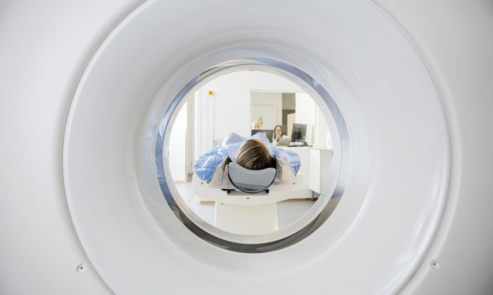
PET/CT scans
Two weeks after completing radiotherapy, seven patients (age range: 6-17) were given whole-body PET/CT scans performed at 1, 72 and 144 hours post-injection. The optimal moment of scanning was found to be 144 hours post-injection. The patients also underwent contrast (gadolinium)-enhanced MRI.
“The results showed that indeed there is considerable heterogeneity in uptake of Zr-89-labeled bevacizumab among patients and within tumours,” Van Dongen points out.
“This non-invasive in vivo quantification of drug distribution and tumour uptake may help to predict therapeutic potential, as well as toxicity, and could help develop strategies for improving drug delivery to tumours.”
Van Dongen adds, “Children with brain tumours and other solid cancers are particularly likely to benefit from molecular drug imaging, as drugs without therapeutic effect–based on a lack of drug-uptake in the tumour–may cause life-long side effects. Molecular drug imaging will open avenues for administering the right drug to the right patient at the most appropriate stage of the disease.”
Related topics
Imaging, In Vivo, Radiotherapy, Research & Development
Related conditions
Cancer
Related organisations
VU University Medical Centre
Related people
Guus van Dongen