

The evolving role of three dimensional in-vitro cell culture techniques in drug discovery
Posted: 31 July 2018 | Gowri Sukumar | No comments yet
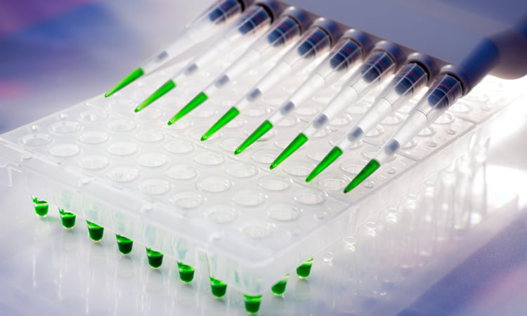
Drug discovery is a lengthy process that proceeds through several stages. High throughput screening (HTS) utilising whole-cell based screening assays play a fundamental role as a starting point for identifying novel compounds in the drug discovery process.1,2
Gowri Sukumar, M.S. RAC
Associate Director, CMC & Regulatory Affairs, ESSA Pharmaceuticals
This two-dimensional (2D) approach to cell-based screening involves utilising a monolayer on a variety of planar substrates and has been the preferred method due to the ease with which the cell monolayers can be induced to proliferate on planar surfaces. Though these are widely utilised to predict in vivo drug responses for many targets and pathways, however, there are challenges with the current 2D model since it does not accurately represent the microenvironment especially for the tumour cells and thus difficulties may be encountered downstream with translation in vivo. The disadvantages are primarily associated with the loss of tissue architecture, mechanical and biochemical cues, and cell-to-cell -to- matrix interactions, thus making them relatively poor models to predict drug responses especially for diseases such as cancer.
Drug Target Review has just announced the launch of its NEW and EXCLUSIVE report examining the evolution of AI and informatics in drug discovery and development.
In this 63 page in-depth report, experts and researchers explore the key benefits of AI and informatics processes, reveal where the challenges lie for the implementation of AI and how they see the use of these technologies streamlining workflows in the future.
Also featured are exclusive interviews with leading scientists from AstraZeneca, Auransa, PolarisQB and Chalmers University of Technology.
Today, the limitations of 2D culture, the advancement of laboratory products, high-end detection instrumentation and analytical software as well emergence of stem cell had led to the growing adaptation of the 3D culture models in, many phases of drug discovery, including target validation, lead identification as well as preclinical optimization. Thus, tissue specific architecture along with elements in surrounding microenvironment, are essential components for in vivo response and can be at least partially recapitulated utilizing Three- Dimensional (3D) cell culture models (3). Previously, 3D culture models were mostly developed for cancer research, given that the environment in the 3D culture more closely mimics the tumor microenvironment. At present, 3D cell culture techniques are emerging as a pivotal new tool in preclinical drug discovery. The past decades have witnessed rapid progress in the development of 3D cell cultures and recent studies have demonstrated the potential of utilizing these culture models in such applications (4,5,6).
There are a wide range of 3D cell culture technologies that have emerged as a result of recent advances in tissue engineering, microfabrication and cell biology. 3D cell tumor models exist in several forms from multicellular layers on porous membranes coated with collagen to matrix-embedded cultures, hollow fiber bioreactors and multicellular spheroids. Recently, technologies from stem cell research as well mixed cell culture techniques have been the key to growing use of dish-based organogenesis (7). This technique helps establish a high degree of physiological relevance alongside facilitating the development of organoids that exhibit a realistic micro-anatomy. This article will provide an overview on the common 3D culture techniques as well as address the advantages and challenges.
3D cell culture techniques:
The term 3D cell culture relates to cell growth in some kind of matrix or scaffold with 3-Dimensional structure which would stimulate a tissue-specific physiological or pathophysiological disease-specific microenvironment whereby the cell can proliferate, aggregate and differentiate (8). There are many approaches and technologies to the cell growth in the 3D cell cultures. The 3D cell culture models are being validated using an array of new assay formulation, multiplexing and staining methods aimed at measuring several parameters such as DNA staining of dead cells, caspase makers for apoptosis and others (9).
Primarily, 3D cell culture systems are classified into two categories; 1. Scaffold- free 2. Scaffold based (natural or synthetic). For scaffold-based 3D cell culture systems, there exists a wide variety of materials that are available for use. As an estimate, over 100 types of matrices (from proteins, glycoproteins to secreted proteins and growth factors) as well as scaffolds (both organic and in-organic) are in use (10). Extracellular matrix (ECM) though was previously thought to serve as a structural support but is now known to play a critical role in various aspects of cellular behaviour in a tissue specific manner. In addition, the level of rigidity of the material used for the 3D cell growth being one of the characteristics that influence the maintenance of homeostasis (11). Therefore, the composition of ECM along with its physical properties can have a major impact on cell’s response to drugs either by enhancing its efficacy, altering its Mechanism of Action (MOA) or by increasing drug resistance (12).
Scaffold- free technologies:
Anchorage-independent 3D culture techniques rely on the self-aggregation of cells on specialized culture plates. There are several variations to the specialized culture plates and include the hanging drop microplates and the low adhesion plates (with ultra-low attachment coating that promotes the formation of spheroids) (13). Hanging drop cultures are a popular 3D technology that takes advantage of self- aggregation of cells into spheroids when the surface is not available for attachment and has applications in toxicity testing in hepatocytes and in engineering cardiac spheroids (14,15). The low adhesion plates are similar to hanging drop cultures since it takes advantage of lack of cell attachment surface to promote aggregation of cells and spheroid formation. However, this technique has other advantages overhanging drop, with the elimination of steps that are required for manipulating spheroids. Another emerging technique for the formation of spheroids is the Magnetic cell levitation which also holds promise in drug discovery (16). This technique employs magnetic nanoparticles that are preloaded on the cells. And then using an externally applied magnetic field, the cells are floated toward the air/liquid interface to promote cell-cell aggregation and thus the facilitate spheroid formation (17).
Scaffold- Based Technologies:
Anchorage-dependent culture techniques provide physical support ranging from simple mechanical structure to ECM like matrices which facilitate aggregation, proliferation and migration of cells. Typically, the cells are embedded into the matrix and the physical and chemical properties of the scaffold material will influence the cell characteristics. Scaffolds of synthetic origin can be modified to have biologically active molecules to be encapsulated in order to enhance specific phenotype or aid in cell proliferation. Therefore, the selection of scaffold is a critical step for a given application with consideration on the properties of the material that define physical as well as biological factors. Scaffolds can be broadly classified into Hydrogel scaffolds of biological origin and synthetic hydrogels. Hydrogels are networks formed from dilute polymer chains with given structure and properties which are formed as a result of intermolecular or interfibrillar crosslinks.
Hydrogel Scaffolds of biological origin from natural sources include collagen, fibrin or Matrigel and are biocompatible with natural adhesive properties and sustain many physiological cell functions. Type I Collagen is the most commonly utilized ECM in 3D culture (18,19). In contrast, synthetic hydrogels which can mimic biological properties of ECM and are classified as non-natural and natural polymers. Unnatural polymer hydrogels include Polyethylene glycol (PEG), polylactic acid (PA), Polyglycolic acid (PGA) and others. These have the advantage of being relatively inexpensive with reproducible material properties, however lack adhesive moieties found in natural ECM (20). Synthetic natural polymers also offer the advantage of consistent and tunable material and properties. In addition, are highly compatible with encapsulation of cells due to their biological nature of their naturally occurring moieties. Hydrogels in Hyaluronic acid (HA) is an example of one of the best characterized natural hydrogels attributing to presence of glycosaminoglycan which can be modified with functional groups, allowing for the formation of hydrogels with an array of properties with diverse applications (21).
As the materials science continues to evolve, a new range of hydrogels like small peptide-based materials will provide new insights to the 3D cell culture technology. Also, Micropatterned plates can be designed to serve as a scaffold-based 3D culture environment with an endless array of imprinted micropatterns on the surface of the plates (22). Microspun 3D fibrous scaffold was recently developed to aid tumoroid formation for anticancer drug development (23).
Despite the major progress in 3D cultures over the past few years offering considerable promise across a wide range of in drug discovery and toxicity testing for a diverse range of diseases from cancer to neurological disorders, this technology has some limitations. Primarily due the inability of this technology to be able to provide relevant and validated 3D cell/ tissue models. The major challenge in the optimization stems from lack of the quantifiable entity of biomarkers of three dimensionalities. On the other hand, there are some limitations that are specific to the method type as well. One such example can be the differences in the reproducibility between different batches for the scaffold- based culture system which is still unsatisfactory. There are also reports of potential interactions of the screening compounds with the scaffold which can in turn impact the quality of the results from the screening studies.
In conclusion, while there are several advantages of 3D cell culture technologies from the perspective of biological relevance, especially for drug discovery and other applications. However, there are still some challenges which need to be addressed to establish 3D culture systems as a standardized platform for drug discovery and other applications in the pharmaceutical industry.
References:
- Swinney DC, Anthony J. Nat. Rev. Drug Discov. 2011, 10(7), 507.
- Swinney DC. Clin. Pharmacol. Ther. 2013, 93(4), 299.
- Benien P, Swami A. Future Oncol. 2014, 10(7), 1311
- Montanez-Sauri SI, Sung KE, Berthier E, Beebe DJ. Enabling screening in 3D microenvironments: probing matrix and stromal effects on the morphology and proliferation of T47D breast carcinoma cells. Biol.2013, 5, 631–640. doi: 10.1039/c3ib20225a
- Ryan SL, Baird AM, Vaz G, Urquhart AJ, Senge M, Richard DJ., et al. Drug discovery approaches utilizing three-dimensional cell culture. Assay Drug Dev. Technol.2016, 14, 19–28. doi: 10.1089/adt.2015.670
- Edmondson R, Broglie JJ, Adcock AF, Yang L. Three-dimensional cell culture systems and their applications in drug discovery and cell-based biosensors. Assay Drug Dev. Technol. 2014, 12, 207–218. doi: 10.1089/adt.2014.573
- Huch, M, Koo, BK. Development, 2015, 142(18), 3113.
- Griffith LG, Swartz MA. Capturing complex 3D tissue physiology in vitro. Rev. Mol. Cell Biol. 2006, 7, 211–224. doi: 10.1038/nrm1858
- Riss TL, Valley MP, Zimprich CA, Niles AL, Kupcho KR, Lazar DF. Design and Validation of Bioluminescent Assays for 3D Cell Culture Models, Promega Corporation poster,2014, Abstract#226
- Hynes RO, Naba A. Overview of the matrisome–an inventory of extracellular matrix constituents and functions. Cold Spring Harb. Perspect. Biol. 2012, 4:a004903. doi: 10.1101/cshperspect.a004903
- Handorf AM, Zhou Y, Halanski MA, Li WJ. Tissue stiffness dictates development, homeostasis, and disease progression. Organogenesis. 2015, 11, 1–15. doi: 10.1080/15476278.2015.1019687
- Bonnans C, Chou J, Werb Z. Remodelling the extracellular matrix in development and disease. Rev. Mol. Cell Biol.2014, 15, 786–801. doi: 10.1038/nrm3904
- Sutherland RM. Cell and environment interactions in tumor microregions: the multicell spheroid model. Science. 1988, 240, 177–184. doi: 10.1126/science.2451290
- Shri M, Agrawal H, Rani P, Singh D, Onteru SK. Hanging drop, a best three-dimensional (3D) culture method for primary buffalo and sheep hepatocytes. Rep. 2017, 7:1203. doi: 10.1038/s41598-017-01355-6
- Chitnis T, Weiner HL.CNS inflammation and neurodegeneration. Clin. Invest.2017, 127, 3577–3587. doi: 10.1172/JCI90609
- Timm DM, Chen J, Sing D, Gage JA, Haisler WL, Neeley SK, et al. A high-throughput three-dimensional cell migration assay for toxicity screening with mobile device-based macroscopic image analysis. Rep. 2013, 3:3000. doi: 10.1038/srep03000
- Haisler WL, Timm DM, Gage JA, Tseng H, Killian TC, Souza GR. Three-dimensional cell culturing by magnetic levitation. Protoc.2013, 8, 1940–1949. doi: 10.1038/nprot.2013.125
- Orgel JP, Persikov AV, Antipova O. Variation in the helical structure of native collagen. PLoS ONE. 2014, 9:e89519. doi: 10.1371/journal.pone.0089519
- Pathak A, Kumar S. Biophysical regulation of tumor cell invasion: moving beyond matrix stiffness. Biol. 2011, 3, 267–278. doi: 10.1039/c0ib00095g
- Kraehenbuehl TP, Zammaretti P, Van der Vlies AJ, Schoenmakers RG, Lutolf MP, Jaconi ME, et al. Three-dimensional extracellular matrix-directed cardioprogenitor differentiation: systematic modulation of a synthetic cell-responsive PEG-hydrogel. 2008, 29, 2757–2766. doi: 10.1016/j.biomaterials.2008.03.016
- Baeva LF, Lyle DB, Rios M, Langone JJ, Lightfoote MM. Different molecular weight hyaluronic acid effects on human macrophage interleukin 1beta production. Biomed. Mater. Res. A. 2014, 102, 305–314. doi: 10.1002/jbm.a.34704
- Knight E, Murray B, Carnachan R, Przyborski S. Alvetex(R): polystyrene scaffold technology for routine three dimensional cell culture. Methods Mol. Biol.2011, 695, 323–340. doi: 10.1007/978-1-60761-984-0_20
- Girard YK, Wang C, Ravi S, Howell MC, Mallela J, Alibrahim M, et al. A 3D fibrous scaffold inducing tumoroids: a platform for anticancer drug development. PLoS ONE8:e75345. 2013, doi: 10.1371/journal.pone.0075345
Related topics
High Throughput Screening (HTS), Hit-to-Lead, Screening