


news
Novel 3D imaging technology could improve fluorescence microscopy
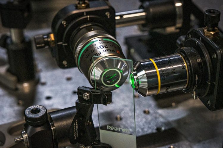
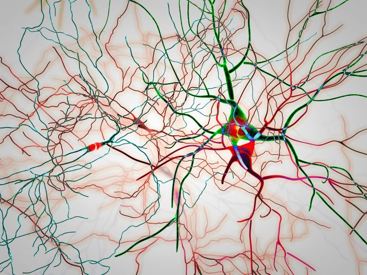
A new technique called Coded Light-sheet Array Microscopy (CLAM) has been developed by researchers to improve 3D imaging of living specimens.
List view / Grid view
A new technique called Coded Light-sheet Array Microscopy (CLAM) has been developed by researchers to improve 3D imaging of living specimens.
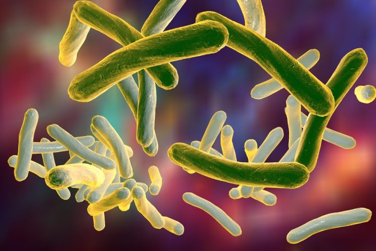

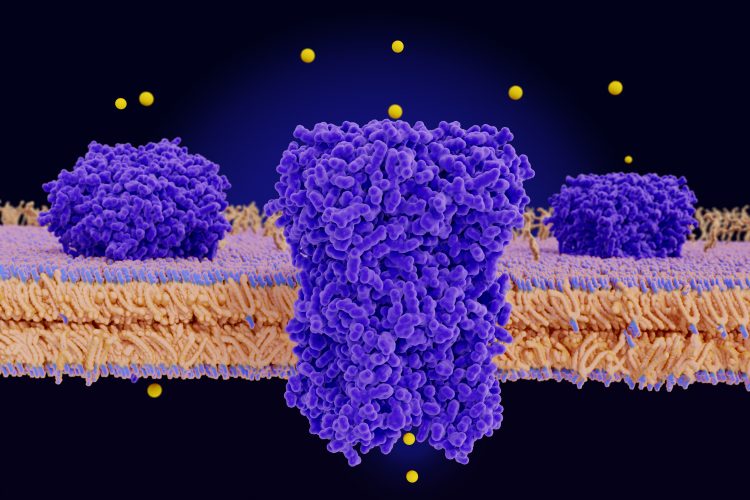
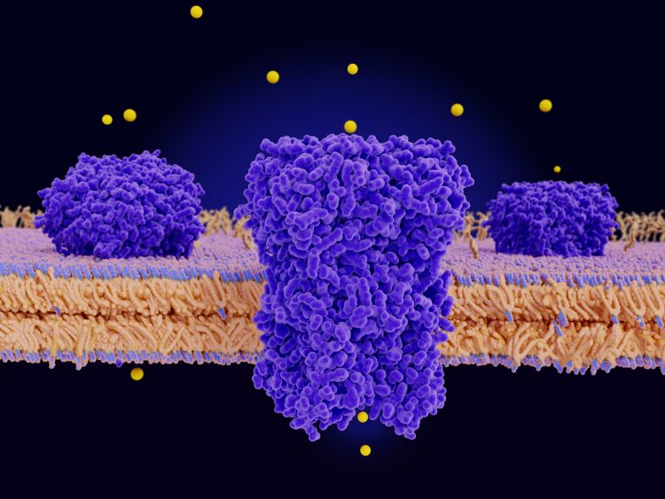
Cryogenic electron microscopy revealed that the vitamin B12 transporter on Mycobacterium tuberculosis acts like a non-selective sluice, transporting both the vitamin and antibiotics.
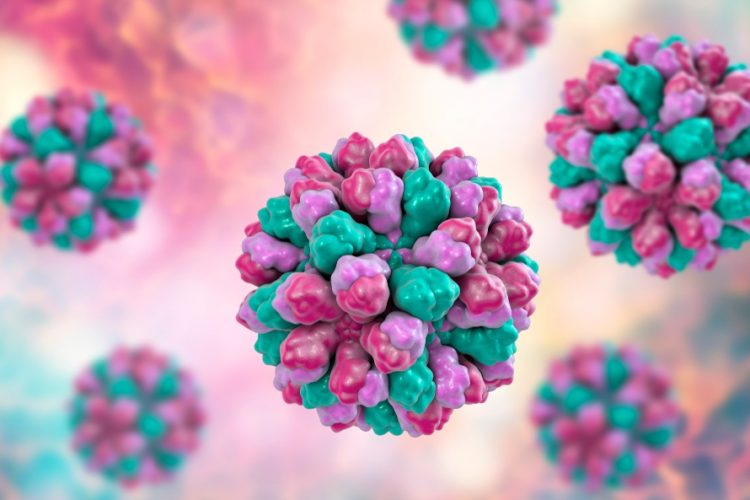
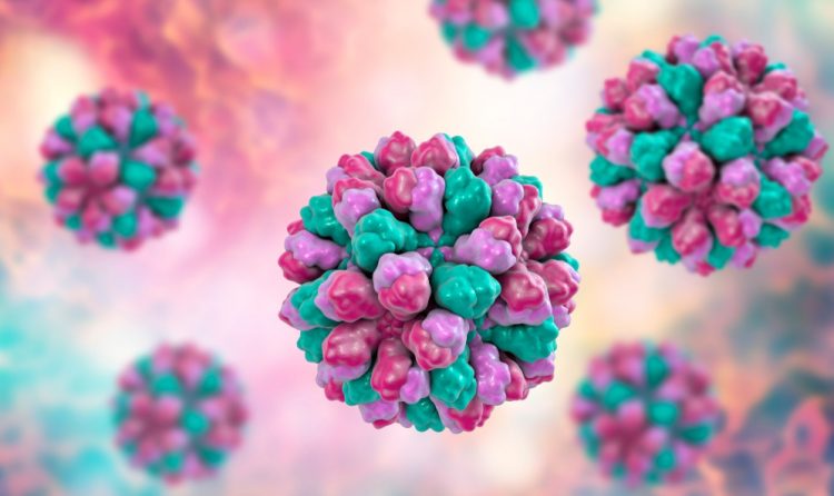
Researchers captured 13,000 images of a mouse norovirus using an electron microscope and compiled the images to reveal the structure of the virus.
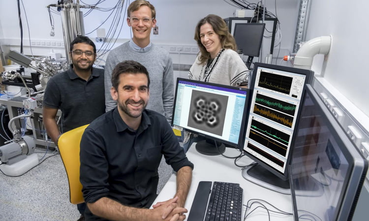
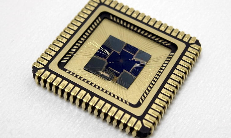
Using AI and deep learning, researchers have enhanced Scanning Probe Microscopy (SPM) and made their automated resource available for scientists.
Scientists have imaged the ball-and-chain mechanism using cryogenic electron microscopy and hope their work could be applied in the design of novel therapeutics.
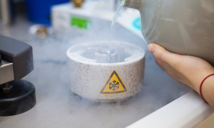
Researchers have created a new cryogenic electron microscopy (cryo-EM) technique by utilising low-energy electrons in a holographic method.
Researchers have identified that copper ions and their protein transporters, such as Atox1, are key to cancer cell movement and could be targeted by therapies.
Using fluorescent markers, researchers have developed Förster resonance energy transfer (FRET) to image the assembly, functions and interactions of molecules.
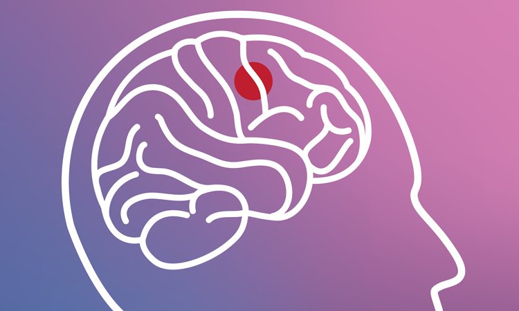
Scientists have identified two master controller regions that are essential for alpha-synuclein aggregation and could be targeted by future therapies.
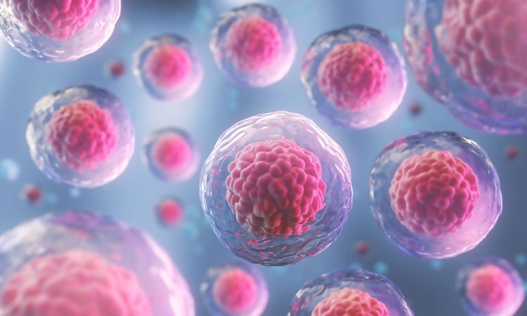
A new method has been created by researchers to 3D bioprint tumours and image glioblastomas for the study of therapeutics.
Researchers have applied for a patent for their innovative cantilever and vibrating plate technique which they say could increase the speed of atomic force microscopy on fragile samples.
A new imaging technique, which has revealed 3D forces exerted by tiny cell clusters, could help scientists understand how tissue forms, how wounds heal or how tumours spread.
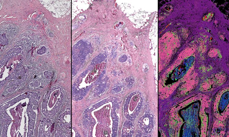
A new method to image cancerous tissues has been created by researchers who have paired infrared measurements with high-resolution optical images.
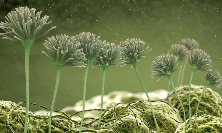
Researchers have studied how the human body responds to viral infection when already infected by fungi, offering insights into the immune system.
Using cryogenic electron microscopy, a team has mapped the Spike protein on COVID-19, which could be used in the development of vaccines.