

news
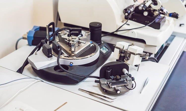
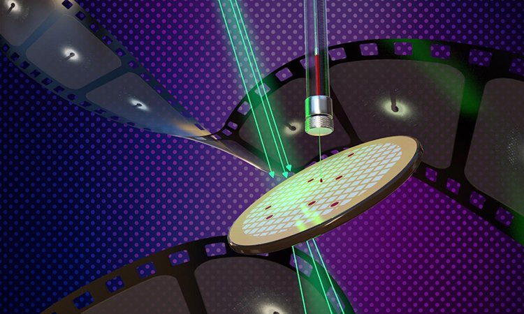
Atomic force microscopy reveals structure of ribosome stalk proteins
Researchers have used force atomic microscopy to show the structural dynamics at ribosome stalk proteins when building new proteins.
List view / Grid view
Researchers have used force atomic microscopy to show the structural dynamics at ribosome stalk proteins when building new proteins.
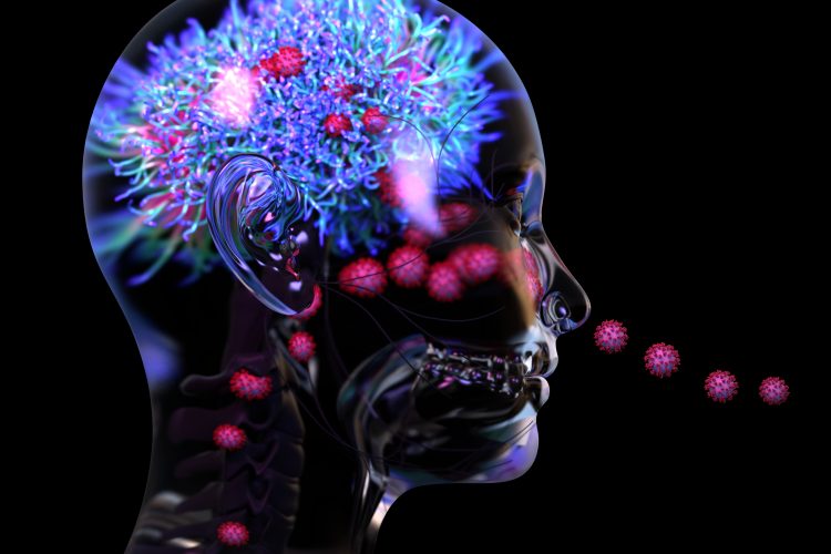
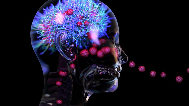
A new study suggests that inflammation and blood vessel damage may be the primary causes of neurological symptoms in COVID-19 patients, instead of the virus infecting the brain.
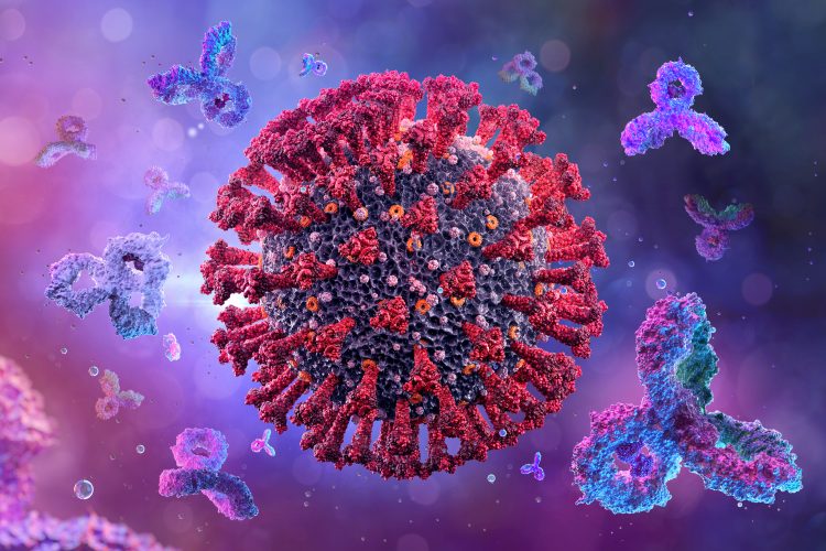
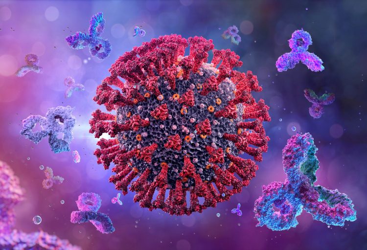
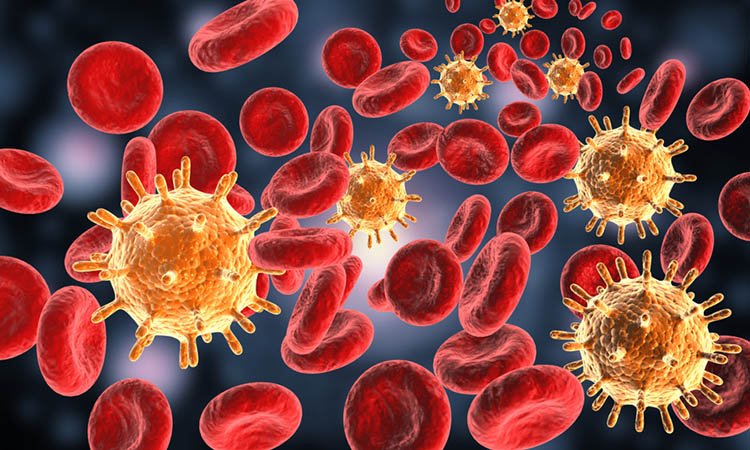
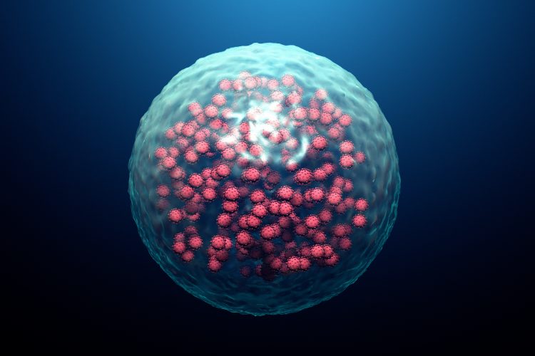
Researchers show that neutralising antibodies targeting the SARS-CoV-2 Spike protein have four distinct structures.
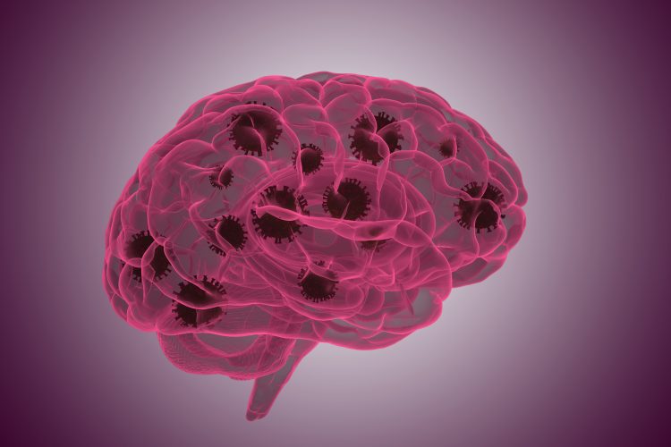
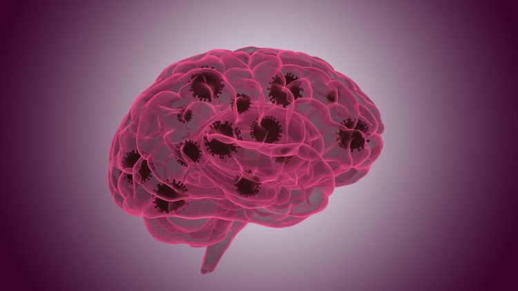
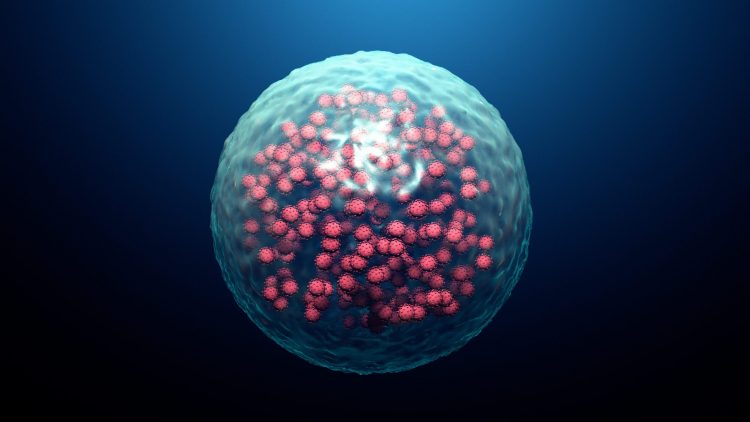
A new study has identified the mechanisms through which the SARS-CoV-2 virus enters the brain and how the immune system responds once it does.
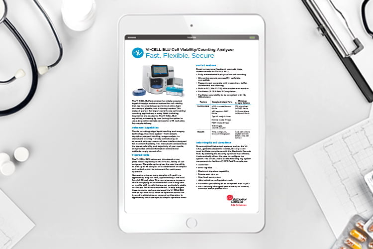
The Vi-CELL BLU automates the widely accepted trypan blue dye exclusion method for cell viability that has historically been performed with a light microscope, pipette, and a hemacytometer.
A new imaging method called FLASH can provide a visualisation of several tissue types in a 3D format, its developers say.
This article lists three of the most recent advances in pre-clinical HIV research and vaccine development.
The study shows how drug-like small molecules inhibit the activity of Transient Receptor Potential Canonical 1/4/5 (TRPC1/4/5) channels and could transform the development of future therapies.
Scientists have shown how SARS-CoV-2 induces changes in the architecture of host cells to drive replication and made their data available to all.
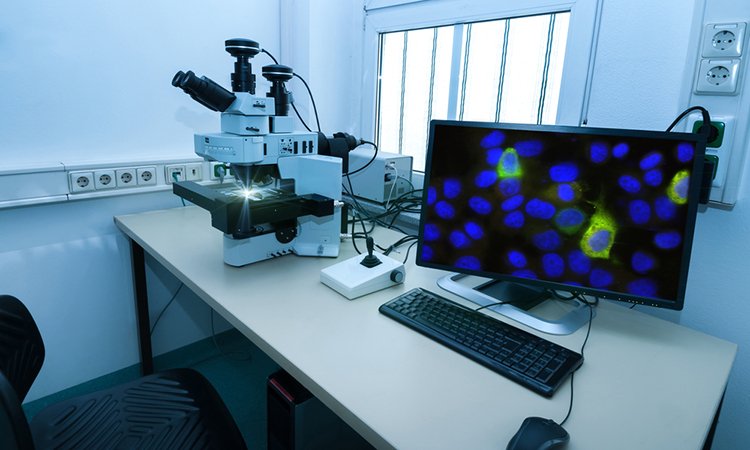
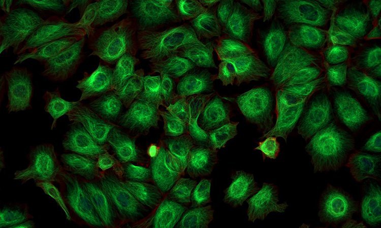
Using a new CRISPR-Cas9 tagging strategy, researchers have developed a method that enables the imaging of hundreds of proteins in parallel.
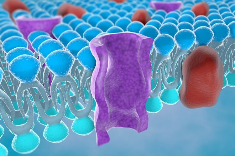
Lan Zhu from Arizona State University explains how cryo-EM methods can be used to obtain structural information on membrane proteins such as GPCRs.

Identify therapeutic effects and adverse responses to compounds earlier in the drug discovery process.
Using cryo-electron microscopy and site-specific mass spectrometry, researchers have mapped the glycans that shield HIV from the immune system.

A major challenge within electrophysiology labs is 50 or 60 Hz line frequency electrical noise, which can either distort or completely drown out biological signal.

Combining 2D and 3D models with live-cell assays allows monitoring of cell responses in real time and provides important insights about compound treatment effects, biological complexity, and physiological relevance of assay results.