

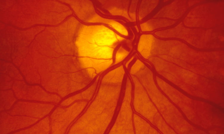
Researchers map pathoconnectome of eye disease and retinal circuitry
Posted: 30 September 2020 | Victoria Rees (Drug Target Review) | No comments yet
Scientists have produced the first pathoconnectome – an open access map for other researchers that shows how eye disease can alter retinal circuitry.
Scientists have achieved a first in the field of connectomics, producing the first pathoconnectome that shows how eye disease alters retinal circuitry. The study was conducted at the John A Moran Eye Center at the University of Utah, US.
Moran’s Marclab for Connectomics was the first to complete a map of the circuitry of the retina, or connectome, in 2011. Now, the US National Institutes of Health (NIH)-funded lab has produced the first pathoconnectome, a map of neural network synaptology and circuit topologies corrupted by neurological disease to identify robust targets for therapeutics.
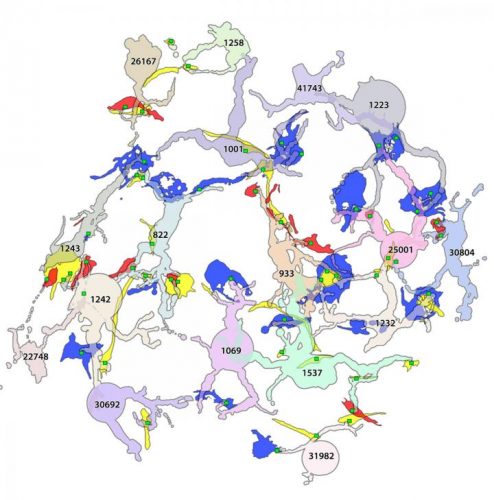
A 2D pathoconnectome image shows rod bipolar cell dendrites and their synapse locations with rod (red), cone (blue) and indeterminate (yellow) photoreceptors [credit: John A Moran Eye Center].
The Marclab developed the pathoconnectome from a model of early-stage retinitis pigmentosa (RP), an inherited retinal disease that can lead to blindness. This pathoconnectome was generated using serial section transmission electron microscopy to achieve an ultrastructural connectome for the identification of all chemical and gap junctional synapses. The researchers say they observed aberrant connectivity in the rod-network pathway and novel synaptic connections deriving from neurite sprouting.
The large dataset compiled to construct the pathoconnectome has taken the team years to assemble and is open for use by other scientists. The Marclab is now working on a second and third pathoconnectome that will show how the retina rewires itself in later stages of RP.
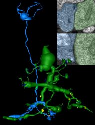
A 2D pathoconnectome image showing two retinal neurons (rod bipolar cell in blue, Aii amacrine cell in green). A gap junction connection (lower inset image) is not normally present between them but formed as the degenerating retina rewired itself during disease [credit: John A Moran Eye Center].
According to the researchers, the implications of this research extend far beyond eye diseases. They say the eye holds lessons applicable to a host of neurodegenerative diseases including Alzheimer’s, Parkinson’s, epilepsy and Lou Gehrig’s disease.
“The components of neurodegeneration we see in the eye seem to mimic those we see in the brain,” explained the paper’s lead author, Dr Rebecca Pfeiffer. “So this pathoconnectome is allowing us to learn fundamental rules of how neurodegenerative diseases alter neural networks in general. The ultimate goal is to identify how we might develop new therapies based on preventing or interfering with the network rewiring prompted by disease.”
The study was published in Experimental Eye Research.
Related topics
Neurons, Neurosciences, Research & Development
Related conditions
Alzheimer's disease (AD), Epilepsy, Lou Gehrig's Disease, Parkinson's disease, Retinitis pigmentosa (RP)
Related organisations
University of Utah, US National Institutes of Health (NIH)
Related people
Dr Rebecca Pfeiffer