

The Olympus scanR high-content screening station rapidly acquires quantitative data from cell-based assays
Posted: 6 December 2019 | Olympus | No comments yet
Self-learning microscopy opens new horizons in high-content analysis and advances phenotypic screening.
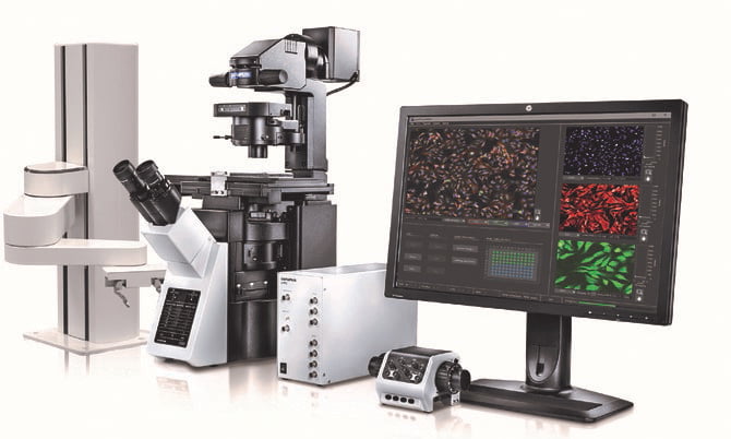
Olympus ScanR Modular High-Content Screening Station for Life Sciences.
Olympus announces the US launch of the scanR high-content screening (HCS) station, a cell imaging solution that utilizes artificial intelligence (AI) to enable next-generation biological research. It combines the modularity and flexibility of a microscope-based setup with the automation, speed, throughput and reproducibility of an HCS station.
AI meets high-content screening
The Olympus scanR HCS station uses the power of AI to carry out groundbreaking analyses of cells that, until recently, seemed impossible to do. Olympus’ self-learning microscopy technology reduces photobleaching and improves acquisition speed, measurement sensitivity, and accuracy, facilitating longer observations with reduced influence on cell viability.
The power of deep learning
The system acquires pairs of images that are processed by the software to generate an image analysis model. Olympus’ optimized deep-learning technology, which is based on a dedicated convolutional neural network architecture, provides powerful and flexible learned analysis protocols. No human data annotations are required, which allows large numbers of examples to be used, enabling the potential of the deep-learning technology to be fully exploited.
Drug Target Review has just announced the launch of its NEW and EXCLUSIVE report examining the evolution of AI and informatics in drug discovery and development.
In this 63 page in-depth report, experts and researchers explore the key benefits of AI and informatics processes, reveal where the challenges lie for the implementation of AI and how they see the use of these technologies streamlining workflows in the future.
Also featured are exclusive interviews with leading scientists from AstraZeneca, Auransa, PolarisQB and Chalmers University of Technology.
Self-learning microscopy
After a one-time training phase, scanR AI enables the system to automatically analyze new data by incorporating the learned analysis protocol into its assay-based workflow. Because the user has full control in designing the training experiment and many challenging analysis conditions can be covered during the training phase, the accuracy and robustness of the analysis results are improved.
Robust, fast and automated image acquisition and analysis

Self-Learning Microscopy: AI prediction of nuclei positions (blue), green fluorescent protein (GFP) histone 2B labels showing nuclei (green), and raw brightfield transmission image (gray).
The scanR system performs fully automated image acquisition and analysis of multiwell plates, slides and custom-built arrays. Throughout the automated image acquisition, the scanR system maintains the focus plane using a combination of software algorithms and hardware, including TruFocus Z-drift compensation. Images are automatically analyzed during acquisition to minimize analysis time, and all units are precisely synchronized by a real-time controller to maximize the acquisition speed.
Simple workflow and fast setup to ease research
The scanR HCS station combines these benefits with a user-friendly workflow that requires less human interaction, even during the AI training stage. The responsive interface contributes to intuitive, straightforward analysis. This makes it easy for life science and pharmaceutical researchers to harness the power of HCS data while benefitting from a fast, simple setup.
For more information, please visit here.
About Olympus
Olympus is a global technology leader, crafting innovative optical and digital solutions in medical technologies; life sciences; industrial solutions; and cameras and audio products. Throughout our 100-year history, Olympus has focused on being true to society and making people’s lives healthier, safer and more fulfilling.
Our Life Sciences Business is dedicated to meeting and exceeding the evolving needs and expectations of life science professionals through a comprehensive range of clinical, educational and research microscopes and microscope systems. For more information, visit https://www.olympus-lifescience.com and truetolife.com.
Olympus…True to You. True to Society. True to LIFE.
Related topics
Artificial Intelligence, High-content assays, Research & Development, Screening, Technology