

First detailed 3D image of astrocytes produced by researchers
Posted: 30 October 2019 | Victoria Rees (Drug Target Review) | No comments yet
Researchers have compiled images to create the first detailed 3D models of astrocytes, which could be used in the development of therapeutics to aid their function.
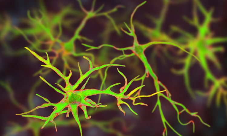
A number of imaging experiments led by researchers at the King Abdullah University of Science and Technology (KAUST), Saudi Arabia, have produced three-dimensional (3D) models of astrocytes.
According to the researchers, their imaging technique will enable scientists to design drugs that aid astrocyte function.
“Astrocytes are cells that store energy and give energy to neurons at multiple levels,” said Dr Corrado Calì, the lead researcher. “To do that, it was important to produce accurate 3D reconstructions of these astrocytes.”
Drug Target Review has just announced the launch of its NEW and EXCLUSIVE report examining the evolution of AI and informatics in drug discovery and development.
In this 63 page in-depth report, experts and researchers explore the key benefits of AI and informatics processes, reveal where the challenges lie for the implementation of AI and how they see the use of these technologies streamlining workflows in the future.
Also featured are exclusive interviews with leading scientists from AstraZeneca, Auransa, PolarisQB and Chalmers University of Technology.
The team used serial block-face electron microscopy to generate an image stack. These were compiled to produce a volumetric image of a piece of tissue that contains astrocytes which could then be segmented.
As astrocytes are extremely complex in structure, it is impossible to image them in their entirety using light or fluorescent techniques, unless using electro-microscopy.

Dr Corrado Calì explores 3D reconstructions of extremely complex brain cells known as astrocytes, a key cell involved in memory formation and learning, within KAUST’s Visualization Core Laboratories (credit: KAUST).
The researchers suggest that their study will help in revealing the full function of astrocytes in the brain. It may also aid in designing drugs that improve the function of these cells to treat pathological conditions such as stroke or Alzheimer’s disease.
“We could help to slow down the disease by acting specifically on these cells,” Dr Calì noted.
The findings were published in Progress in Neurobiology.
Related topics
Drug Targets, Imaging, Neurons, Neurosciences, Research & Development
Related conditions
Alzheimer’s disease, Stroke
Related organisations
King Abdullah University of Science and Technology (KAUST)
Related people
Dr Corrado Calì