

Non-invasive imaging for small airway lung disease
Posted: 14 March 2019 | Iqra Farooq (Drug Target Review) | No comments yet
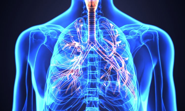
A non-invasive method of imaging tiny bronchioles in the lungs of chronic obstructive pulmonary disease means patients could detect the disease earlier…
Chronic obstructive pulmonary disease (COPD) is the fourth leading cause of death in the US, with approximately 16 million Americans currently affected.
Researchers at the University of Michigan have reported on the ability of Parametric Response Mapping (PRM), a relatively new technique, to identify small airway abnormality in COPD.
Currently, it is difficult to identify abnormalities in small airways non-invasively, as the tiny bronchioles that are initially damaged are around two millimetres in internal diameter, and as such are too small to be imaged using computed tomography.
Drug Target Review has just announced the launch of its NEW and EXCLUSIVE report examining the evolution of AI and informatics in drug discovery and development.
In this 63 page in-depth report, experts and researchers explore the key benefits of AI and informatics processes, reveal where the challenges lie for the implementation of AI and how they see the use of these technologies streamlining workflows in the future.
Also featured are exclusive interviews with leading scientists from AstraZeneca, Auransa, PolarisQB and Chalmers University of Technology.
PRM was developed at Michigan Medicine, in a study led by Dr Brian Ross, Professor of Radiology and Biological Chemistry, and Dr Craig Galban, Associate Professor of Radiology. The technique measures lung density during inhalation and exhalation.
Using lung tissue from patients undergoing transplantation, and donated healthy lung tissue, researchers mapped the samples back to CT scans taken before surgery. The team was able to non-invasively identify small airway loss, narrowing and obstruction.
Senior author Dr MeiLan Han, a lung specialist and Professor of Internal Medicine at the University of Michigan, said, “Now we have confidence in our ability to identify airway disease when imaging COPD patients. PRM is already clinically available and used by University of Michigan clinical teams to assess patients with COPD. This is what we mean by bench to bedside medicine.”
The study was conducted in patients with a severe form of the disease. A NHLBI funded study, COPDGene, found that the PRM-defined small airway abnormalities have been detected on CT scans of patients with milder disease and could help to predict patients who will lose lung function.
Prof Han said, “We still need to validate the type of airway disease the PRM technique identifies in patients with milder disease. That type of lung tissue is more difficult to obtain, but we are working on techniques that would allow us to use smaller amounts of lung tissue to make such studies feasible.”
The study was published in the American Journal of Respiratory and Critical Care Medicine.
Related conditions
COPD
Related organisations
Michigan University
Related people
Dr Brian Ross, Dr Craig Galban, Dr MeiLan Han