

Researchers develop biosensor with real-time oxygen monitoring
Posted: 21 August 2018 | Iqra Farooq (Drug Target Review) | No comments yet
In a study, researchers have developed a biosensor concept using organs-on-a-chip to measure the levels of oxygen in real-time…
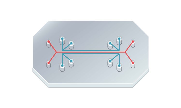
Researchers have developed a method using biosensors to track oxygen levels in real time in ‘organ-on-a-chip’ systems. This makes it possible for the system to closely mimic the function of actual organs and is essential for the system to be successful in drug and toxicity testing.
The concept of an organ-on-a-chip is to create small-scale biological structures that mimic the function of a specific organ, for example transferring oxygen from the air into the bloodstream, in a manner similar to that of lungs.
Also called microphysiological models, they are used to expediate high-throughput testing, to evaluate the effectiveness of a new drug or to assess toxicity. Despite the increased advances in this technique over the last decade, one obstacle to its progress is the lack of tools designed to actually retrieve the data.
Drug Target Review has just announced the launch of its NEW and EXCLUSIVE report examining the evolution of AI and informatics in drug discovery and development.
In this 63 page in-depth report, experts and researchers explore the key benefits of AI and informatics processes, reveal where the challenges lie for the implementation of AI and how they see the use of these technologies streamlining workflows in the future.
Also featured are exclusive interviews with leading scientists from AstraZeneca, Auransa, PolarisQB and Chalmers University of Technology.
“For the most part, the only existing ways of collecting data on what’s happening in an organ-on-a-chip are to conduct a bioassay, histology, or use some other technique that involves destroying the tissue,” said Assistant Professor Michael Daniele, from North Carolina State University and corresponding author of the paper.
“What we really need are tools that provide a means to collect data in real time without affecting the system’s operation,” aid Prof Daniele. “That would enable us to collect and analyse data continuously and offer richer insights into what’s going on. Our new biosensor does exactly that, at least for oxygen levels.”
The levels of oxygen in the body varies widely. In the lungs of a healthy adult, the oxygen concentration would be around 15 percent, whereas in the inner lining of the intestine the level of oxygen would be closer to 0 percent. The level of oxygen directly affects the function of the tissue, and as such when testing using organ-on-a-chip, levels of oxygen need to be realistic.
“What this means in practical terms is that we need a way to monitor oxygen levels not only in the organ-on-a-chip’s immediate environment, but in the organ-on-a-chip’s tissue itself,” Prof Daniele said. “And we need to be able to do it in real time. Now we have a way to do that.”
The researchers developed a phosphorescent gel that emits infrared light after being exposed to infrared light. They describe the process as an ‘echoing flash’, with the time between the gel being exposed to the light and it emitting the light varying, depending on the amount of oxygen in its environment. The more oxygen present, the shorter the lag time.
Despite the lag time being mere microseconds, the differences between them can be measured and analysed, and oxygen levels can then be measured to one tenth of a percent.
For the biosensor to work, a thin layer of the gel must be incorporated during the manufacture of the organ-on-a-chip. The researchers can use a ‘reader’ to emit the infrared light and measure the echoing flash. Thus, lag times and oxygen levels can be measured.
The team have successfully used this technique in three-dimensional scaffolds using human breast epithelial cells to model cancerous and healthy cells.
“One of our next steps is to incorporate the biosensor into a system that automatically makes adjustments to maintain the desired oxygen concentration in the organ-on-a-chip,” Prof Daniele said. “We’re also hoping to work with other tissue engineering researchers and industry. We think our biosensor could be a valuable instrument for helping to advance the development of organs-on-a-chip as viable research tools.”
Related topics
Drug Discovery Processes, Organ-on-a-Chip, Research & Development, Screening
Related organisations
North Carolina State University
Related people
Assistant Professor Michael Daniele