

Imaging technique to aid IVF embryo selection
Posted: 30 August 2017 | Dr Zara Kassam (Drug Target Review) | No comments yet
Researchers have successfully developed an advanced new imaging technique, which can help assess the quality of early-stage embryos…
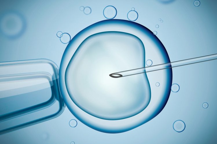
Researchers have successfully developed an advanced new imaging technique, which can help assess the quality of early-stage embryos, this ‘hyperspectral imaging’ measures light that cells naturally produce during their normal activities.
The light or ‘autoflorescence’ produced changes according to the chemical reactions or metabolism going on in the cell. Being able to measure embryo metabolism is viewed by many researchers as one of the most important factors as to whether a particular IVF program will be successful.
“We use a special type of imaging to show differences in the metabolism and chemical make-up of embryos before they’ve been implanted,” says lead author Dr Mel Sutton-McDowall, from the University of Adelaide. “This technique can give us an objective measure of which embryo to choose as part of the IVF process.”
“Pre-implantation screening of embryos generally takes place under a normal optical microscope. Although it’s quite easy to discern poor embryos, it is far harder for the clinician to determine objectively, the viability of the other embryos,” she added. “The challenge is how to choose the single healthiest embryo out of this group to maximise the chances of pregnancy.”
Drug Target Review has just announced the launch of its NEW and EXCLUSIVE report examining the evolution of AI and informatics in drug discovery and development.
In this 63 page in-depth report, experts and researchers explore the key benefits of AI and informatics processes, reveal where the challenges lie for the implementation of AI and how they see the use of these technologies streamlining workflows in the future.
Also featured are exclusive interviews with leading scientists from AstraZeneca, Auransa, PolarisQB and Chalmers University of Technology.
Dr Sutton-McDowall sees the use of hyperspectral imaging as a new tool that can be combined with other diagnostic methods to provide a more accurate and objective embryo viability assessment.
“The benefit of hyperspectral imaging is that it can capture information-rich content of inspected objects. It analyses every pixel in an image for its light intensity at differing wavelengths,” said Dr Sutton-McDowall,.
“This lets us drill down and analyse the hyperspectral signature of each individual embryo, looking for known or anomalous characteristics. It lets us discriminate between embryos, but also measuring metabolic differences within individual embryos. We predict that embryos that have cells with homogeneous metabolic profiles are the healthier ones.”
To date, this imaging technology has only been tested on cattle embryos but is extremely promising. “It offers benefits of being a non-invasive imaging approach that provides real-time information to the clinician,” said Dr Sutton-McDowall,.
The likely development of a specialised hyperspectral imaging tool for actual use in the IVF clinic is several years away but there is a strong surge of interest from IVF clinics to better predict embryo development outcomes through technology.
“IVF is a costly and complex treatment. Any new method that can help improve the odds of women successfully having babies is of benefit to both clinicians and their patients.” Not just limited to human IVF practice, there is also commercial opportunities for the hyperspectral technology across the farming, animal and livestock sectors as well.
The research, reported in the journal Human Reproduction has the potential to significantly benefit the IVF industry of the future, improving assisted reproduction outcomes for women.
Related topics
Imaging, Research & Development
Related organisations
Adelaide University
Related people
Dr Mel Sutton-McDowall