

Power of radiomics to improve precision medicine
Posted: 4 August 2017 | Dr Zara Kassam (Drug Target Review) | No comments yet
Data mining of medical images with radiomics offers clinicians a way to characterise lung tumours and improve precision medicine…
Data mining of medical images with radiomics offers clinicians a way to characterise lung tumours and improve precision medicine.
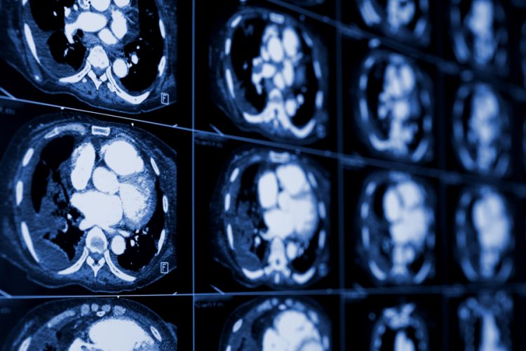
Researchers analysed computed tomography (CT) image features from 262 North American patients and 89 European patients with non-small cell lung cancer (NSCLC). They identified associations between the image features and molecular markers, biological pathways, and clinical outcomes. They determined that certain sets of image features could predict the overall survival of NSCLC patients, while other image features could predict the stage of the tumour or the presence of biological and genetic markers that drive tumour growth.
The researchers also demonstrated the clinical importance of radiomics by showing that it is possible to increase prognostic power by combining radiomic data with genetic information and clinical data.
Drug Target Review has just announced the launch of its NEW and EXCLUSIVE report examining the evolution of AI and informatics in drug discovery and development.
In this 63 page in-depth report, experts and researchers explore the key benefits of AI and informatics processes, reveal where the challenges lie for the implementation of AI and how they see the use of these technologies streamlining workflows in the future.
Also featured are exclusive interviews with leading scientists from AstraZeneca, Auransa, PolarisQB and Chalmers University of Technology.
“The core belief of radiomics is that images aren’t pictures, they’re data. We have to treat them as data. Right now, we extract about 1300 different quantitative features from any volume of interest,” said Dr Robert Gillies, chair of Moffitt’s Department of Cancer Imaging and Metabolism.
“We already knew that radiomic algorithms have strong clinical importance; however, the biological basis for these observations remained unknown. This study now answers this key question for the first time by defining and independently validating the driving biological pathways of radiomic phenotypes” said Dr Hugo Aerts, director of the Computational Imaging and Bioinformatics Laboratory and associate professor of Radiation Oncology at Harvard Medical School.
Radiomics has several advantages over other commonly used techniques that guide precision medicine. Currently, biological markers are routinely analysed with tissue biopsies that are invasive, collected only at the beginning of care, and may not accurately reflect the biology of the entire tumour. In contrast, imaging techniques are non-invasive and can provide information about the entire tumour throughout the entire course of treatment and response. Additionally, the majority of cancer patients routinely have images taken for diagnostic purposes already, making radiomics a cost-effective approach.
“This study advances the molecular knowledge of radiomic characterisation of tumours, information currently not used clinically. This may provide opportunities to improve decision-support in all patients as imaging is routinely used in clinical practice as standard of care,” said Dr Gillies.
Related topics
Big Data, Computerised tomography (CT), Imaging
Related conditions
Lung cancer, non-small cell lung cancer
Related organisations
Harvard Medical School, Moffitt's Department of Cancer Imaging and Metabolism.
Related people
Dr Hugo Aerts, Dr Robert Gillies