

Intercontinental consortium granted €5.1 million to develop breast cancer imaging devices
Posted: 15 September 2016 | Niamh Louise Marriott, Digital Content Producer | No comments yet
Grants of more than €5 million, predominantly from the EU, will be used to develop a new imaging device for the diagnosis of breast cancer…
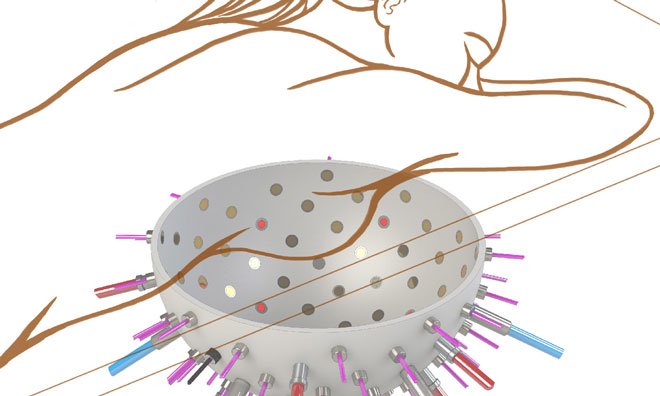
A large European research consortium headed by the University of Twente is to receive grants of more than €5 million, predominantly from the EU, to develop a new imaging device for the diagnosis of breast cancer. A prototype of the device will be ready for large scale testing and production in four years. Not only will it provide improved photoacoustic and ultrasound imaging, it will also be able to combine the images generated by both techniques. The expectation is that the new imager will improve and accelerate the diagnosis of breast cancer, and also be applicable for younger women.
Worldwide, every year more than 1.5 million women are diagnosed with breast cancer; half a million women die from the disease.
Current techniques for detecting breast cancer – x-ray mammography, ultrasound and magnetic resonance imaging (MRI) – have shortcomings.
Drug Target Review has just announced the launch of its NEW and EXCLUSIVE report examining the evolution of AI and informatics in drug discovery and development.
In this 63 page in-depth report, experts and researchers explore the key benefits of AI and informatics processes, reveal where the challenges lie for the implementation of AI and how they see the use of these technologies streamlining workflows in the future.
Also featured are exclusive interviews with leading scientists from AstraZeneca, Auransa, PolarisQB and Chalmers University of Technology.
The most significant disadvantage of these techniques is that they cannot always clearly distinguish a tumour from healthy tissue or a benign abnormality, so that tumours are missed, and often unnecessary – and stressful – biopsies have to be carried out.
A large European research consortium will therefore develop a completely new device for the diagnosis of breast cancer. The proposed device is expected to shorten the time to accurate diagnosis substantially. It will also be suitable for younger women, where x-ray mammography usually does not perform well. The device does not use radiation or contrast media which are potentially harmful, and does not cause any pain to the woman being examined.
The consortium is to receive a grant of €4.35 million from the European Union and the rest from the Swiss government, for the development of the device. Project leader Srirang Manohar calls the proposed device, which has been dubbed ‘PAMMOTH’, a ‘dream imager’ commenting, “we will be working on this prototype with the best partners in Europe and actively involving doctors and patient associations for their inputs and advice in the design and testing processes.”
Combination
Pammoth will combine existing photoacoustic and ultrasound imaging techniques, and elevate both to a higher level at the same time. Manohar explains, “The images from the two systems will be combined together. This will result in simultaneous three-dimensional information about disease specific optical contrast, as well as about the ultrasound properties which provide anatomic information within the breast. Further we wish to do this combined imaging in real time.”
The improvement of both systems will, incidentally, not only be useful for the final clinical prototype on which the researchers will be working. Manohar expects the project to lead to various improved subsystems for data acquisition, ultrasound detection and a new powerful laser.
Photoacoustics
Although photoacoustics is a relatively new imaging technique in the medical world, the UT has been conducting research in its application in breast imaging, which is called photoacoustic mammography or PAMmography, for some time now.
In PAMmography, the breast is illuminated with laser light
In PAMmography, the breast is illuminated with laser light. This laser light is converted into ultrasound in regions where higher concentrations of blood are present, such as the areas around malignant tumours. The ultrasound produced travels from the tumour to the surface of the skin, where it can be detected. The partners participating in the Pammoth project will improve the technique and introduce various novel functionalities.
For example, various colours of laser light will be used to improve visualisation of the tumour, as well as, to yield information about the oxygen saturation of the blood in tumours, which can indicate whether they are malignant or benign.
Ultrasound
The researchers are also hoping to take significant steps in the field of ultrasound. They will develop technology which will yield a three-dimensional image, with the ultrasound waves being generated in an unconventional manner. Contrary to regular ultrasound imaging, hand-held scanners will not be used in this project so as to images from being affected by the operator of the device.
Research consortium
The research consortium will be coordinated by Srirang Manohar from the Biomedical Photonic Imaging group part of the UT research institute MIRA. Besides coordination, the UT will focus on the photoacoustic imaging part, and will conduct research for generating ultrasound for the ultrasound subsystem.
PA Imaging, a UT spin-off company, will develop rapid, low-noise electronics for the detectors and will ensure that the various partners’ components are integrated into the functioning clinical prototype. At the end of the project, Medical Spectrum Twente (MST) will, together with the University of Twente, conduct a pilot study to test, the performance of the system in imaging the most common breast tumours.
Researchers from University College London will work on image reconstruction methods, and will be responsible for the mathematics and calculations required to compute the images. One of the proposed methods will make possible quantitative image reconstruction. The developed mathematics will be converted into usable and rapid algorithms to enable real-time imaging at the Brno University of Technology.
Scientists from the University of Bern will have a role in the analysis of ultrasound images, and will develop specialised breast phantoms from materials having the correct acoustic and photoacoustic properties for testing the system.
The French company Imasonic will design highly sensitive multi-element detectors for detecting the ultrasound, the Lithuanian company Ekspla will develop the lasers needed for the system, and the German company TP21 will be responsible for the management of the project, and the requisite internal information infrastructure.
Horizon 2020
The project will be financed by the European Union’s Horizon 2020 programme which has reserved a total of €80 billion for research and innovation between 2014 and 2020. Essential criteria for eligibility for funding by the programme are excellent scientific research, cooperation between different scientific disciplines and with industry, and whether the research addresses large social problems.
Related topics
Imaging, PA Imaging, TP21
Related conditions
Breast cancer
Related organisations
European Commission (EU), Imasonic, Medical Spectrum Twente (MST), University College London, University of Bern