

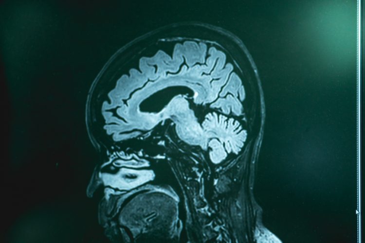
Getting “under the skin” of frontotemporal dementia
Posted: 23 August 2021 | Anna Begley (Drug Target Review) | No comments yet
Researchers at the University of East Finland have been using skin cells to investigate pathological hallmarks in frontotemporal dementia patients.
Frontotemporal dementia (FTD) is the second most common cause of dementia in the working age population, yet it is one of the least understood forms of the disease. Currently, there are no efficient therapies for FTD, it is challenging to diagnose and the disease mechanisms remain largely unclear. What is known, however, is that the most common genetic cause of this dementia is C9orf72 hexanucleotide repeat expansion.
A new study from the University of Eastern Finland aims to tackle the array of research-related problems by investigating whether skin cells from FTD patients show specific cellular pathological hallmarks or functional alterations compared to healthy individuals. According to the researchers, their findings could promote better understanding of molecular mechanisms of FTD and be useful in the discovery of novel biomarkers or in testing drug effects. Both C9orf72 repeat expansion carriers and patients with sporadic FTD, for whom the underlying cause of disease is unknown, were included in the study.
The centrality of neurons
Previous studies on this form of dementia have been scarce as cellular pathological changes related to the C9orf72 repeat expansion have not been widely described in other cells than neurons so far. In an exclusive interview with Drug Target Review’s Anna Begley, Research Director Annakaisa Haapasalo explained that while there are some studies on other cells, research has largely focused on neurons as they are the principal central nervous system (CNS) cells that are affected in neurodegeneration.
Drug Target Review has just announced the launch of its NEW and EXCLUSIVE report examining the evolution of AI and informatics in drug discovery and development.
In this 63 page in-depth report, experts and researchers explore the key benefits of AI and informatics processes, reveal where the challenges lie for the implementation of AI and how they see the use of these technologies streamlining workflows in the future.
Also featured are exclusive interviews with leading scientists from AstraZeneca, Auransa, PolarisQB and Chalmers University of Technology.
“Current neuropathological studies have indicated that the C9orf72 repeat expansion-associated pathologies, such as RNA foci and dipeptide repeat proteins as well as C9orf72 haploinsufficiency, are most prevalently present in neurons as compared to other brain cells,” explained Haapasalo. “Therefore, it is natural that their effects on neurons are under intensive study and neurons are also one of our key research interests.”
Using skin cells to understand FTD
However, Haapasalo and her team proposed an alternative approach: studying skin cells. “We became interested in exploring the FTD patient skin fibroblasts in more detail with the idea in mind that they might represent more easily accessible patient-derived cells than neurons for trying to decipher underlying disease mechanisms,” revealed Haapasalo. “We were interested in finding out if these cells show any specific alterations or deficiencies that could be utilised later on in biomarker studies or testing drug effects.”
We became interested in exploring the FTD patient skin fibroblasts in more detail with the idea in mind that they might represent more easily accessible patient-derived cells than neurons for trying to decipher underlying disease mechanisms.”
The team obtained skin biopsy samples from FTD patients through clinical collaborators from Kuopio University Hospital and established the fibroblast cultures from these biopsy samples in the laboratory. They then used a range of biochemical and molecular biological methods, fluorescence microscopy and live-cell assays to characterise and assess the function and properties of these cells.
The researchers’ findings indicate that skin fibroblasts of carriers of the C9orf72 expansion partially display similar pathological changes to those found in the brain. Patient skin cell cultures may therefore possess potential as platforms for testing drug effects when screening compounds that could prevent formation of the abnormal RNA foci and the subsequent pathological dipeptide repeat (DPR) proteins derived from these abnormal RNAs.
The role of p62 in skin fibroblasts
One of the researches’ most promising findings was the significantly increased accumulation of vesicles containing a protein known as p62 in the skin fibroblasts of both sporadic and C9orf72 expansion-carrying FTD patients. Accumulation of p62 may be a sign of defective ability of the cells to degrade proteins, however defects in the function of the main cellular protein degradation routes, the proteasomes or autophagosomes, were not detected in this study. The findings raise the question of whether the increased number and size of p62 vesicles in skin fibroblasts could be utilised as disease biomarkers for diagnosing this type of dementia.
This discovery is particularly exciting as it could lead to further novel discoveries and Haapasalo indicated that the role of p62 may be an area of focus in the team’s future research. “We would like to investigate further if the accumulation of p62 vesicles is specific only to FTD patient fibroblasts or if it is detectable also in patients with other neurodegenerative diseases,” she said. “If it was FTD patient-specific, it is possible that it might be employed as a biomarker for these patients but clarifying this will need further investigations.”
Investigating mitochondrial energy metabolism
Haapasalo noted that another pivotal finding was the defective mitochondrial energy metabolism in the FTD patient fibroblasts when compared to the fibroblasts from healthy donors. The skin fibroblasts from both sporadic and C9orf72 expansion-carrying FTD patients displayed a significantly weaker energy metabolism. These changes were detected in assays where the basal respiration and ATP-mediated energy production by the cells’ power plants, the mitochondria, were measured.
“What was interesting was that these changes were not detected only in the fibroblasts of the C9orf72 repeat expansion carriers but also in fibroblasts from sporadic FTD patients,” said Haapasalo. “They appear to be changes detectable in FTD patients’ fibroblasts in general and not linked specifically to the C9orf72 repeat expansion. It might be interesting to assess next whether the cellular changes could be utilised in biomarker studies or as endpoints in, for example, the testing of drug effects.”
The endless possibilities of skin cells
One of the significant advantages of the study is that the changes observed in the skin fibroblasts are partially similar to those observed in the brain of FTD patients. “Because brain cells can rarely be obtained from the brains of living patients, other patient-derived cells, such as skin fibroblasts, are extremely useful in research,” commented Haapasalo. “Their use enables us to clarify disease mechanisms at the cellular and molecular level and may prove useful in biomarker or drug research, even at the individual level.”
In addition, patient-derived skin cells could be utilised as sources to produce induced pluripotent stem cells (iPSCs). These cells can be further differentiated in the laboratory to different types of brain cells and used as human disease models in research, Haapasalo added. Meanwhile in her lab, FTD patient-derived skin cells have been used to generate iPSCs that were further differentiated into different types of brain cells, such as neurons and microglia.
The study on dementia was published in Molecular Neurobiology.
Related topics
Biomarkers, Cell culture, DNA, Genetic analysis, Genomics, In Vitro, Molecular Targets, Neurons, Neurosciences, Protein, Protein Expression, RNAs, Target molecule, Targets
Related conditions
frontotemporal dementia (FTD)
Related organisations
University of Eastern Finland
Related people
Annakaisa Haapasalo