

Protecting liposomes in COVID-19 mRNA vaccines could increase their stability
Posted: 27 April 2021 | Victoria Rees (Drug Target Review) | No comments yet
A team from the University of Texas at Dallas has shown that a novel buffer solution is effective at stabilising the liposomes and lipid nanoparticles in certain COVID-19 vaccines.
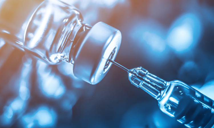
New research findings have the potential to address a major challenge in the deployment of certain COVID-19 vaccines worldwide – the need for the vaccines to be kept at below-freezing temperatures during transport and storage.
With regular news updates and feature articles, our COVID-19 hub has everything you need to keep up to date with R&D during the pandemic. Click below to visit:
The study, conducted at the University of Texas at Dallas, US, revealed that a new, inexpensive technique can generate crystalline exoskeletons around liposomes and other lipid nanoparticles to stabilise them at room temperature for up to two months.
Published in Nature Communications, the researchers say the study provides a proof-of-concept to develop the formulation of certain of COVID-19 vaccines.
Drug Target Review has just announced the launch of its NEW and EXCLUSIVE report examining the evolution of AI and informatics in drug discovery and development.
In this 63 page in-depth report, experts and researchers explore the key benefits of AI and informatics processes, reveal where the challenges lie for the implementation of AI and how they see the use of these technologies streamlining workflows in the future.
Also featured are exclusive interviews with leading scientists from AstraZeneca, Auransa, PolarisQB and Chalmers University of Technology.
The need for specialised storage requirements
According to the study researchers, the Pfizer/BioNTech and Moderna COVID-19 vaccines use lipid nanoparticles – spheres of fat molecules – to protect and deliver the messenger RNA (mRNA) that generates a vaccine recipient’s immune response to the SARS-CoV-2 virus, by providing the instructions to build a structure similar to the coronavirus’ Spike (S) protein. The body’s immune response is then triggered, meaning that neutralising antibodies are generated and will be again upon infection from SARS-CoV-2.
“The expense of keeping these vaccines very cold from the time they are made to the time they are delivered is a challenge that needs to be addressed, especially because many countries do not have sufficient infrastructure to maintain this kind of cold chain,” said Dr Jeremiah Gassensmith, a corresponding author of the study. “Although we did not include in this work the specific lipid nanoparticles used in current COVID-19 vaccines, our findings are a step toward stabilising a lipid nanoparticle in a way that has never been done before, so far as we know.”
The idea for the research project began during a discussion between Gassensmith and Dr Gabriele Meloni, a corresponding co-author of the study. Gassensmith’s area of expertise is biomaterials and metal-organic frameworks, while Meloni’s research focus is transmembrane transporter proteins. These proteins reside within cell membranes and are crucial for moving a variety of small molecules, including ions and trace metals, in and out of cells for several purposes.
“Membrane proteins sit in a cell membrane, which is a lipid bilayer,” Meloni said. “To study their structure and biophysical and biochemical properties, we must extract these proteins from the membrane using detergents and then reconstitute them back into an artificial membrane – a proteoliposome – that mimics the proteins’ natural environment.”
Creation of a shell

University of Texas at Dallas scientists developed a method to stabilise liposomes in a crystalline exoskeleton, which allows the biomolecules to remain stable at room temperature. This illustration depicts a proteoliposome – a spherical bilayer of fat molecules (white and blue) – stabilised in a structure called a zeolitic-imidazole framework composed of zinc acetate and methylimidazole. Inserted into the lipid bilayer – which mimics a cell membrane – are modelled structures of CopA proteins, with a section (in pink) that resides inside the lipid and sections above the lipid surface (brown) and slightly inside the liposome (also brown, but inside) [credit: University of Texas at Dallas].
Lipid nanoparticles and liposomes are similar in structure and neither are thermodynamically stable at room temperature, Gassensmith said. The lipid structures can fuse or aggregate, exposing any embedded membrane proteins or cargo to degradation.
“One of the challenges in my field of research is that both membrane proteins and lipid bilayers are very delicate and intrinsically metastable and we are trying to combine them in order to understand how these proteins function,” Meloni said. “We have to handle them carefully and prepare them fresh each time. They cannot be stored for long periods and are not easily shipped to colleagues in other labs.”
The researchers joined forces to develop a methodology capable of stabilising this kind of vaccine lipid system. They demonstrated their results using transmembrane proteins from Meloni’s lab as a case study.
They mixed liposomes – some with embedded proteins, some without – with a combination of two inexpensive chemicals, zinc acetate and methylimidazole, in a buffer solution. Within approximately one minute, the team saw that a crystal matrix began to form around individual liposomes.
“We think that the lipids interact with the zinc just strongly enough to form an initial zinc-methylimidazole structure that then grows around the lipid sphere and completely envelops it, like an exoskeleton,” Gassensmith said. “It is analogous to biomineralisation, which is how certain animals form shells. We sort of co-opted nature in creating this totally fake shell, where the biomacromolecules – the lipids and proteins – catalyse the growth of this exoskeleton.”
The ability of biomimetic shells to form around biological molecules is not new, the researchers emphasise, but the process has not worked well with lipids or liposomes before because the metal salts that comprise the shell material suck water from the liposomes by osmosis and cause them to explode.
“One of the keys to this research was identifying the buffer solution in which everything resides,” Gassensmith said.
Developing the buffer
Three graduate students collaborated on the project to develop the unique buffer medium that allows the reaction to occur.
“The buffer medium maintains the ionic strength of the solution and keeps the pH stable so that when you add a huge amount of metal salts, it does not osmotically shock the system,” said Fabián Castro, a lead author of the study.
Once the biomolecules have grown a shell, the team explained that they are locked in and the lipids remain stable. While the exoskeleton is very stable, it has a fortuitous weak spot.
“The shell will dissolve if it encounters something that is attracted to zinc,” Gassensmith said. “So, to release and reconstitute the liposomes, we used a zinc chelating factor called ethylenediaminetetraacetic acid (EDTA), which is a common, inexpensive food additive and medicine used to treat lead poisoning.”
 In addition to the laboratory experiments, in another proof-of-concept exercise, Gassensmith mailed through the US Postal Service a sample of the stabilised lipid particles to Rhode Island. These were then shipped back to Texas, but because the COVID-19 pandemic forced the shutdown of many University of Texas at Dallas research labs in 2020, the vaccine samples were untouched for approximately two months after which time the graduate students returned to campus to examine them. Although the informal experiment lasted far longer than the researchers had anticipated, the samples survived and functioned as though they had been stored normally.
“This project required two different types of expertise – my group’s expertise in membrane transport proteins and Gassensmith’s long track record working with metal-organic frameworks,” Meloni said. “Our success clearly demonstrates how such collaborative research can bring about novel and useful results.”
The researchers concluded that this technique could therefore be used to protect and deliver the mRNA in certain COVID-19 vaccines, making them easier to store and transport as the fight against COVID-19 continues.
Related topics
Drug Development, Formulation, Lipidomics, Lipids, Vaccine
Related conditions
Covid-19
Related organisations
BioNTech, Moderna, Pfizer, University of Texas at Dallas
Related people
Dr Gabriele Meloni, Dr Jeremiah Gassensmith, Fabián Castro