Nipah and Hendra virus attack neutralised by potent antibody
Posted: 30 September 2019 | Rachael Harper (Drug Target Review) | 1 comment
Researchers have shown that a monoclonal antibody prevents viruses from fusing with cell membranes to gain entry.


Studies into a new monoclonal antibody (mAb) could pave the way toward preventing Nipah virus and Hendra virus infections or provide a solution for post-exposure therapy, researchers have said.
The team showed that the mAb impedes the fusion machinery henipaviruses use to merge with the membrane of cells they are attempting to breach. It halts the attack by blocking membrane fusion and the injection of the viral genome into the host cell.
The studies were headed by senior researchers David Veesler of the University of Washington School of Medicine’s Department of Biochemistry and Christopher Broder of the Uniformed Services University’s Department of Microbiology and Immunology, both US.
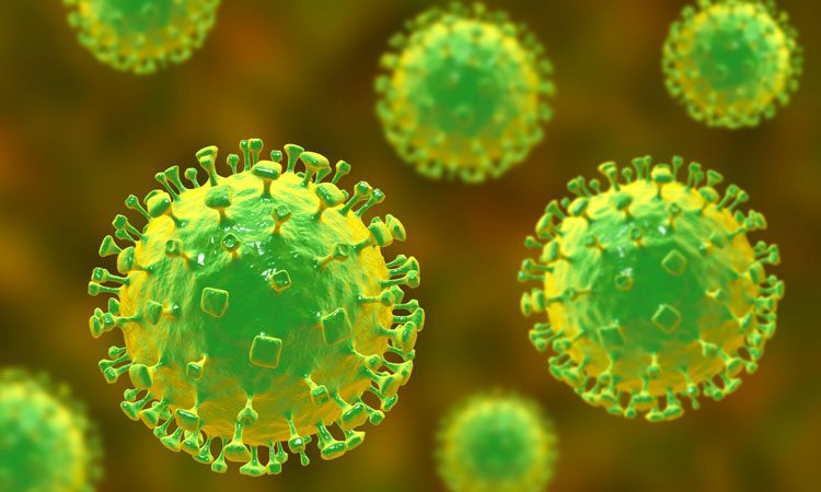
Nipah and Hendra are RNA viruses in the family of paramyxoviruses, many of which have spikey envelopes. The spikes sport a viral-attachment glycoprotein and a fusion glycoprotein. Nipah and Hendra viruses enter cells through the concerted action of the attachment and fusion glycoproteins, the researchers explained. After the virus lands on the cell surface, the attachment protein undergoes a shape change that prompts the fusion protein to insert a fusion peptide into the cell membrane.
…the antibody recognises and binds to a specific area of the viral membrane fusion machinery during the pre-fusion stage.”
Afterwards, the fusion protein refolds into a more stable conformation that staples together the viral and cellular membranes. A tunnel-like pore is forged for the virus’ genetic material to slip into the cell interior to initiate infection. The body’s own humoral immune defenses, mediated by antibody molecules, target vital parts of the merging machinery to keep the virus at bay.
In this latest study, scientists isolated and then humanised from mice, a potent mAb that neutralises both Nipah and Hendra viruses. They then studied its mode of action using molecular imaging via cryo-electron microscopy and biochemical and cellular experiments.
The studies revealed that the antibody recognises and binds to a specific area of the viral membrane fusion machinery during the pre-fusion stage.
This epitope (an antibody target site on the virus) is present in both the Nipah and the Hendra viruses. This explains why the same antibody protects against both viruses. Binding of the antibody prevented membrane fusion and thereby kept the viral material out of the cell.
The report was published in Nature Structural & Molecular Biology.
Related topics
Antibodies, Disease research, Imaging, Monoclonal Antibody, Research & Development, Targets
Related conditions
Hendra virus, Nipah virus
Related organisations
Uniformed Services University, University of Washington School of Medicine
Related people
Christopher Broder, David Veesler


Are the Nipah virus and Hendra virus the same as corona SARs and COVID-19 viruses? Can the monoclonal antibody (mAb) be used on SAR’s or COVID-19 viruses?